


23-month-old female with cough, rhinorrhea, and increased work of breathing
Can you guess the diagnosis?


The case
A 23-month-old female presented with a 3-day history of cough, rhinorrhea, and increased work of breathing. Initially, she was treated for bronchiolitis at an urgent care center, where a nasopharyngeal swab identified adenovirus. Despite initial treatment, the patient's condition worsened, and she required oxygen supplementation via nasal cannula for oxyhemoglobin desaturation. Her hypoxemia further deteriorated, necessitating an increase in oxygen supplementation (Figure 1).
On physical examination, the patient showed signs of respiratory distress. A chest X-ray (CXR) revealed opacities in the right middle and lower lobes. The patient's condition improved with supportive care, and she was discharged after several days in the hospital.
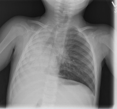
However, 9 months later, due to recurrent wheezing, she was referred to the pulmonary service for further evaluation. A follow-up CXR, taken 3 months after the initial infection, demonstrated complete opacification of the right hemithorax with a mediastinal shift to the right as well as collapse of the right upper and middle lobes.


Evaluation
On physical examination, the patient appeared in mild respiratory distress. Her oxygen saturation was 96% on room air. Breath sounds were diminished on the left side, with crackles noted in the right lower lung fields. Her growth and development were normal, and she was in the 25th percentile for both height and weight. A complete blood count and metabolic panel were within normal limits.


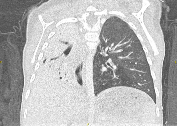
 and (B):
Axial and coronal chest CT views 3 months after the patient's initial infection demonstrate pronounced mediastinal shift with a presumed collapse of the right upper and middle lobes. There is dense airspace consolidation throughout the lower right lobe with superimposed bronchiectasis.
Click image to zoom in")
Due to recurrent wheezing, she was referred to the pulmonary service. A follow-up CXR (3 months afterward) showed complete opacification of the right hemithorax with mediastinal shift to the right and collapse of the right upper and middle lobes (Figure 2). CT scan of the chest revealed unilateral hyperlucency of the left lung. There was complete opacification of the right hemithorax, mediastinal shift to the right, and atelectasis of the right upper and middle lobes with superimposed bronchiectasis (Figures 3A and B). Her findings were consistent with Swyer-James-Macleod Syndrome (SJMS) (Table). Her condition was managed with inhaled corticosteroids, chest physiotherapy, and antibiotics during exacerbations. Her most recent CXR, 15 months after initial presentation, continues to reveal hyperlucency of the left lung (Figure 4).


Management
Her condition was managed with inhaled corticosteroids, chest physiotherapy, and antibiotics during exacerbations.


Discussion
We present a rare case of SJMS in a pediatric patient. SJMS is a postinfectious bronchiolitis first described by Swyer and James in 1953.1-4 This condition is characterized by unilateral hyperlucency of the lung, typically following a viral or bacterial infection in infancy or early childhood. Our patient’s diagnosis followed recurrent respiratory infections and lung collapse.
SJMS can be associated with numerous infections, including adenovirus types 3, 7, and 21, paramyxovirus, Bordetella pertussis, mycobacteria, Mycoplasma pneumoniae, influenza virus, Streptococcus pneumoniae, and Staphylococcus aureus.1 These infections lead to inflammation and fibrosis that cause bronchial lumen narrowing, reduced ventilation/perfusion, and vasoconstriction, consequently reducing blood flow to the pulmonary artery and leading to arterial hypoplasia.5 In addition, decreased pulmonary vascularization, abnormal alveolar development, and destruction of the pulmonary parenchyma and alveoli are observed.6
Due to its rarity, little is known about the incidence of SJMS, and the clinical course varies based on the presence or absence of bronchiectasis.5 Adults with SJMS are often asymptomatic, with the condition being an incidental finding on imaging. In contrast, children tend to be symptomatic, presenting in infancy or early childhood with dyspnea, wheezing, recurrent respiratory infections, productive cough, failure to thrive, and, rarely, hemoptysis and spontaneous pneumothorax.2,5,6
Diagnostic tools such as bronchoalveolar lavage have shown active inflammatory processes in SJMS, with increased neutrophils and lymphocytes, specifically CD8+ T cells.8 Pulmonary function tests may reveal mixed obstructive-restrictive ventilation defects.7 Imaging remains key to diagnosis, with chest x-ray and CT scans revealing characteristic findings of unilateral hyperlucency, bronchiectasis, and lung collapse, as seen in our patient.7
In the differential diagnosis of unilateral hyperlucency (Table), conditions such as congenital lobar emphysema (CLE), congenital pulmonary airway malformation, and pulmonary sequestration must be considered. CLE presents with respiratory distress in infancy and hyperinflation of the lung lobe, whereas SJMS typically follows an infectious process and affects an entire lung. Pulmonary sequestration, involving nonfunctioning lung tissue with an aberrant blood supply, also needs to be ruled out using imaging studies such as CT or MRI.
Treatment for SJMS is preventative and conservative, with a focus on averting further infection. Management includes airway clearance with chest physiotherapy, vaccinations against influenza and pneumococcal infections, and antibiotic treatment during exacerbations.2,7 The efficacy of surgical interventions, such as lung resection, is not well established but remains an option in cases of clinical pulmonary deterioration, recurrent infections despite prophylactic measures, or recurrent pneumothoraces.8 Further studies are needed to explore the benefits of inhaled medications, including bronchodilators and inhaled corticosteroids with or without long-acting beta agonists.
This case highlights the importance of recognizing SJMS as a sequela of early childhood respiratory infections. Early diagnosis and intervention can help reduce the long-term risk of complications such as bronchiectasis and progressive lung disease.
References
- Wojcicki KM, Sindel AD, Berry AC, et al. 2015. An uncommon obliterative lung disease: Swyer‐James‐MacLeod syndrome. Intern Emerg Med. 2015;10:881-882. doi:10.1007/s11739-015-1239-z
- Chaucer B, Chevenon M, Toro C, et al. 2015. Swyer‐James‐MacLeod syndrome: a rare finding and important differential in the ED setting. Am J Emerg Med. 2016;34(7):1329.e3-4. doi:10.1016/j.ajem.2015.12.045
- Kawasuji H, Suzuki K, Furuse H, Tsuda T, Masaki Y, Taniguchi H. Multimodal imaging findings in an adult case of Swyer-James-MacLeod syndrome. Respirol Case Rep. 2017;5(4):e00236. doi:10.1002/rcr2.236
- MacLeod WM. Abnormal transradiancy of one lung. Thorax. 1954;9:147-153.
- Vieira Gde D, Yamagishi AY, Vieira NN, Fogaça RM, Alves Tda C, Amaral GM, Sousa CM. Complication of post-infectious bronchiolitis obliterans (Swyer-James syndrome). Rev Assoc Med Bras (1992). 2015;61(5):404-406. doi:10.1590/1806-9282.61.05.404
- Pérez Guerrero JJ, Sánchez Salguero CA. Hiperclaridad pulmonar unilateral en un niño. Síndrome de Swyer-James-Macleod. Article in Spanish. Arch Argent Pediatr. 2019;117(5):e527-e531. doi:10.5546/aap.2019.e527
- Mehra S, Basnayake T, Falhammar H, Heraganahally S, Tripathi S. Swyer-James-MacLeod syndrome-a rare diagnosis presented through two adult patients. Respirol Case Rep. 2017;5(5):e00245. doi:10.1002/rcr2.245
- Mariana M, Gaio R, Albuquerque J, Gonçalves M, Lobo L. Swyer-James-Macleod Syndrome presentating as pneumothorax. J Ped Surg Case Rep. 2018;37:57-59. doi:10.1016/j.epsc.2018.07.015
Newsletter
Access practical, evidence-based guidance to support better care for our youngest patients. Join our email list for the latest clinical updates.