Boy’s progressive extremity rash looks fishy
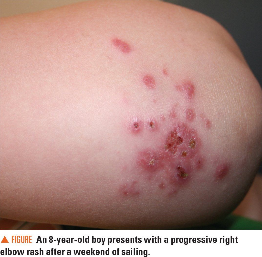
A previously healthy 8-year-old boy presents to the dermatology clinic with a progressively worsening elbow rash over the course of the last week. The rash does not itch. He spent the previous weekend sailing on the Chesapeake Bay. His pediatrician prescribed a course of cephalexin as well as a trial of topical antiviral ointment, neither of which improved the rash. The patient denies any other new exposures.
Figure

The case
A previously healthy 8-year-old boy presents to the dermatology clinic with a progressively worsening elbow rash over the course of the last week. The rash does not itch. He spent the previous weekend sailing on the Chesapeake Bay. His pediatrician prescribed a course of cephalexin as well as a trial of topical antiviral ointment, neither of which improved the rash. The patient denies any other new exposures.
Diagnosis: Mycobacterium marinum infection
Discussion
Mycobacterium marinum is a slow-growing, nontuberculous photochromogen found in a variety of bodies of water.1 Also known as “swimming pool granuloma” or “fish tank granuloma,” cutaneous M marinum infections are most often reported among fish fanciers and those who participate in waterborne activities.2
Mycobacterium marinum disease in humans is uncommon, with rates of up to 0.27 cases per 100,000 persons noted in the literature.2 Although cutaneous M marinum on the limbs is the most common form of the infection, invasive disease (tenosynovitis, arthritis, osteomyelitis) also can occur. The incubation period is usually within 4 weeks following trauma to the skin but can be up to 9 months.
Lesions resulting from cutaneous M marinum infection can present as nodules, ulcers, abscesses, and/or pustules (Figure).1
Data on M marinum susceptibility are limited.1 Biopsy is the preferred diagnostic modality. Unlike rapid-growing mycobacteria, M marinum usually requires several weeks before sufficient culture growth occurs.3 In this patient, granulomas were seen upon microscopic review of the biopsy sample, and culture eventually grew M marinum.
Management
For moderate cutaneous M marinum disease in pediatric patients, the following antibiotics can be considered: rifampin, trimethoprim-sulfamethoxazole, clarithromycin, or doxycycline.3 Extensive lesions may require surgical debridement. Susceptibility testing is not routinely required.
In one case series from France, adults with cutaneous infections were treated with a median duration of 4 months of antibiotics.1 Patients with cutaneous infection in this series mostly received monotherapy with clarithromycin or a cycline. Deeper structure infections were often treated with combination antibiotic therapy with regimens including clarithromycin, rifampin and/or ethambutol. Of the 63 patients studied, 30 with cutaneous and/or deeper structure infections underwent surgery for excision and/or debridement.
Another retrospective case series from Duke University noted that boating or fishing exposure seemed to be associated with invasive disease, whereas fish tank exposure was associated with cutaneous disease.2 In this series, median time to diagnosis for invasive infection was 4 months. This delay in diagnosis indicates that the indolent nature of invasive M marinum infection requires a high index of suspicion in those with aquatic exposure.
Of note, patients with a nontuberculous mycobacterium infection can have positive subsequent tuberculin test results because of shared antigens between M tuberculin-derived proteins and proteins from other nontuberculous species.3 Interferon-gamma release assays also may give positive results attributed to antigen cross-reactivity.
Patient outcome
The patient was treated with a 3-month course of trimethoprim-sulfamethoxazole, based on culture susceptibility testing. His lesions healed fully within 1 to 2 years.
References:
1. Aubry A, Chosidow O, Caumes E, Robert J, Cambau E. Sixty-three cases of Mycobacterium marinum infection: clinical features, treatment, and antibiotic susceptibility of causative isolates. Arch Intern Med. 2002;162(15):1746-1752.
2. Johnson MG, Stout JE. Twenty-eight cases of Mycobacterium marinum infection: retrospective case series and literature review. Infection. 2015;43(6):655-662.
3. Diseases caused by nontuberculous Mycobacteria (environmental Mycobacteria, Mycobacteria other than Mycobacterium tuberculosis. In: Baker CJ, ed. Red Book Atlas of Pediatric Infectious Diseases. 3rd ed. Elk Grove Village, IL: American Academy of Pediatrics; 2016:686-688.

Recognize & Refer: Hemangiomas in pediatrics
July 17th 2019Contemporary Pediatrics sits down exclusively with Sheila Fallon Friedlander, MD, a professor dermatology and pediatrics, to discuss the one key condition for which she believes community pediatricians should be especially aware-hemangiomas.