History of Cough in an Infant and a Toddler
A 6-month-old boy with 1-week history of dry cough that worsened at night. He had been wheezing off and on for the past month and had visited the emergency department on one occasion.

CASE 1 | HISTORY
A 6-month-old boy with 1-week history of dry cough that worsened at night. He had been wheezing off and on for the past month and had visited the emergency department on one occasion. He had also vomited 4 times in the past 24 hours but had been drinking and eating well. He had no fever. His 2-year-old sister was also sick with cough and diarrhea. Infant lived with an aunt who smoked.
Birth history included mild neonatal jaundice that resolved and a small ventricular septal defect and patent foramen ovale that have been monitored by cardiology.
PHYSICAL EXAMINATION
Temperature was 36.5ºC (97.7ºF); heart rate, 120 beats per minute; respiration rate, 40 breaths per minute with moderate distress; capillary oxygen saturation, 97% on room air. Length at the 10th percentile, weight between the 10th and 25th percentile. Eyes, tympanic membranes, and pharynx normal. Mucous membranes moist and tears present. Chest examination demonstrated subcostal and intercostal retractions. Auscultation revealed good air exchange bilaterally with no rales, but with diffuse wheezing and a grade 2 early systolic ejection murmur over the precordium. Rapid respiratory syncytial virus (RSV) test results negative.

CASE 2 | HISTORY
A 2-year-old girl with severe nighttime cough, congestion, and fever (temperature up to 39.5ºC [103ºF]) for the past 2 days. She had recently recovered from a respiratory tract illness of 1 month’s duration. She had been treated with nebulized albuterol, oral prednisolone, and azithromycin. Mother was frustrated because of the child’s persistent illness. Child also had occasional cough with exertion and lived with her parents who claimed to smoke outside the house.
PHYSICAL EXAMINATION
Temperature was 38.9ºC (102.1ºF); heart rate, 95 beats per minute; respiration rate, 38 breaths per minute with considerable distress; capillary oxygen saturation, 87% to 89% on room air. Growth parameters normal. Chest examination demonstrated suprasternal, intercostal, and subcostal retractions with diffuse wheezing and rhonchi. Rapid RSV and influenza test results negative.
DIAGNOSTIC IMAGING
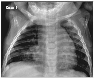
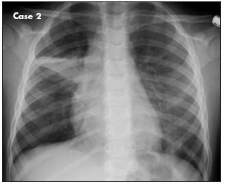
Posteroanterior chest radiographs of both patients are shown.
WHAT'S YOUR DIAGNOSIS?
WHAT'S YOUR DIAGNOSIS?
ANSWER: REACTIVE AIRWAY DISEASE

RADIOGRAPHIC FINDINGSCase 1. The chest radiograph, which is slightly rotated as evidenced by the clavicles, shows a normal heart contour with an upper border merging with thymic tissue, a right middle lobe infiltrate, and peribronchial cuffing in both lung fields. A lateral film (not shown) revealed flattening of the diaphragm, suggestive of hyperinflation and/or air trapping.
Although congestive heart failure might be considered on the basis of the patient’s symptoms, the intermittent wheezing, stable cardiac lesions, and acute pulmonary infiltrate favor a diagnosis of recurrent respiratory illness, manifested as acute bronchiolitis and right middle lobe pneumonia.

Case 2. The chest radiograph, which is minimally rotated, shows right middle lobe atelectasis, supported by a slight rightward shift of the mediastinum and mild hyperinflation in the upper and lower lung fields bilaterally. Persistent wheezing with right middle lobe atelectasis is suggestive of asthma.
RECOGNITION OF RECURRENT RESPIRATORY DISEASE
Reactive airway disease, whether transient or persistent, is one of the most common pediatric complaints.1 The essential symptom of wheezing may appear insidiously at night with accompanying cough, after exercise (which in infants can include crying or excitement), or with respiratory distress and fever that reflect bronchiolitis or pneumonia or both. Pediatricians must recognize the initial symptoms of wheezing, tachypnea, and/or cough; document their recurrence and frequency; and eliminate other causes before making a diagnosis of asthma.
INCIDENCE
About 12.7% of children receive an asthma diagnosis some time before age 18 years; this amounts to 6 million children in the United States.1 Despite the advent of remarkably effective therapies and protocols,2 asthma still causes 7 million ambulatory visits; 200,000 hospitalizations; and 13 million missed school days each year.
ETIOLOGY
Reactive airway disease has many causes of which asthma, defined by atopy and entwined with allergic rhinitis and eczema, is the most common.3 A history of prematurity favors lung injury (bronchopulmonary dysplasia), and association with eating or ingestion favors aspiration or a foreign body. Infection with a virus (eg, RSV) or bacteria (eg, Mycobacterium tuberculosis, pneumococcus) can produce transient wheezing; in this setting, patient age determines whether pneumonitis presents as bronchiolitis or cough.
Congenital anomalies of the trachea or pulmonary tree must be suspected in children with birth defect patterns, and wheezing caused by dysphagia and aspiration should be suspected in those with global delays. Singlegene disorders, such as cystic fibrosis, often have associated symptoms and familial cases that reveal the diagnosis. Similarly, a family history of atopy supports the diagnosis of asthma and draws attention to the “allergic march” of eczema/allergic rhinitis/asthma that can be slowed by effective therapy.
Closely intertwined with asthma is bronchiolitis- the most common lower respiratory tract disease in young children. In children younger than 2 years, bronchiolitis is defined by rhinorrhea, cough, wheezing, tachypnea, and increased respiratory effort (grunting, nasal flaring, intercostal/subcostal retractions).4,5 RSV infection is a major contributor to these reactive airway diseases, infecting 90% of children younger than 2 years, with 40% of infections involving the lower respiratory tract. RSV infection causes 90,000 hospitalizations per year in the United States and reinfections can occur throughout life.
Symptoms of bronchiolitis with a negative result on a rapid test for RSV-as in Case 1-identifies a child at risk for recurrent respiratory disease. A diagnosis of bronchiolitis rather than asthma is important because bronchodilator therapies are not recommended for bronchiolitis unless a positive response to this therapy has been previously documented for the patient by objective measures (eg, oxygen saturation, flow parameters).4-6
Evidence of recurrence and findings associated with asthma (atopy by skin testing, positive family history) are needed to determine whether an episode of bronchiolitis foretells a diagnosis of asthma.
DIAGNOSIS
Accurate diagnosis is important to differentiate asthma from disorders that may not respond well to asthma therapy. The latter include allergic rhinitis, gastroesophageal reflux disease, cardiac failure, laryngotracheomalacia, and vocal cord dysfunction.1
A retrospective review of 50 children younger than 3 years evaluated for recurrent respiratory symptoms showed that 20% had wheezing, 12% had wheezing and cough, 11% had cough alone, and 7% had other symptoms (such as, “hard-breathing,” rhinorrhea, or retractions).3 Asthma was diagnosed in 35 of the 50 children, sinusitis in 5, aspiration in 2, and recurrent croup in 1; 7 had no definitive diagnosis at the time of chart review. Those with a diagnosis of asthma were more likely to have recurrent otitis (23 patients), worsening symptoms after exercise (21), a family history of asthma (14), and/or a mother who smoked (10); the patients with asthma were also more likely to snore (11) or have coexisting eczema (7).
The 50 children underwent 1 or more of the following: chest radiography, sweat chloride test, allergy skin test, sinus radiography, and pulmonary function testing/ bronchoscopy.3 Initial chest radiographs often revealed infiltrates during acute attacks; however, findings from subsequent films were normal (with some peribronchial cuffing or perihilar densities), except in children with chronic aspiration. None of the 50 children had pulmonary anomalies, although vascular rings and cystic adenoid malformations have been reported in patients with recurrent respiratory symptoms. In another study, chest radiographs documented asthma findings (hyperinflation with increased lucency, flattened diaphragms, horizontal ribs, increased anteroposterior diameter of the chest) in 94% of 371 patients who had first asthma attacks; 20 of these patients (6%) also had segmental atelectasis and/or pneumonia.6
Children with suspected reactive airway disease need thorough inspection and chest examination for affect (playful vs ill-appearing), posture (relaxed vs upward straining to expand the chest), nasal flaring/obstruction, breath sounds (wheezing with prolonged expiration vs stridor with prolonged inspiration, presence of rales and rhonchi, overall intensity with alertness for silent areas dull to percussion), and respiratory distress (suprasternal, intercostal, and subcostal retractions). Monitor objective data, such as respiration rate and capillary oxygen saturation, as a function of therapy; obtain chest radiographs when warranted by the child’s age, ill appearance, degree of distress/tachypnea, or accessory findings and history (prior rapid progression/recurrence, RSV positivity, poor therapy response). Radiographic findings, including hyperinflation (flattened diaphragms, increased anteroposterior chest diameter, decreased density), peribronchial cuffing, and/or atelectasis, will attest to the bronchiolar involvement of obstructive airway disease and show infiltrates that may suggest coexisting bacterial pneumonia.
However, even with the help that radiographic evidence and other test results can provide, diagnosing the cause of wheezing in children younger than 3 years remains “one of the last true art forms in medicine”3-one that requires broad thinking, the ability to gain the trust and respect of families, and persistence in ruling out possible diagnoses.3
Both of these patients have onset of wheezing at an early age, and their histories could indicate intermittent or mild to moderate persistent asthma at presentation. Additional follow-up is needed to determine whether wheezing episodes persist up to age 6 years to justify the diagnosis of asthma. Parental diaries are useful to document recurrent wheezing episodes, along with their severity and response to therapy regimens.
ACUTE TREATMENT AND OUTCOMECase 1. Acute therapy in this 6-month-old boy involved intramuscular ceftriaxone (75 mg/kg) and home nebulizer treatments with albuterol (1.25 mg every 4 hours). The next day, he presented with minimal respiratory distress. A second injection of ceftriaxone was administered; the parents were instructed to continue oral therapy with cefdinir (250 mg/d) and albuterol treatments for 2 days and to bring the child back if symptoms of fever, wheezing, or respiratory distress developed.
This infant was not given an asthma diagnosis or detailed asthma education/preventive care pending general pediatric follow-up to determine the recurrence of wheezing. The frequency of bronchiolitis in reaction to viral or bacterial infection for children younger than 2 years mitigates the likelihood of recurrent respiratory illness in this child. This conclusion is supported by his prompt response to antibiotics and bronchodilators.
Case 2. Acute therapy in this 2-year-old girl involved continued treatment with nebulized albuterol (1.25 mg every 4 hours) in the hospital plus intravenous prednisolone (2 mg/kg/d) and oral cefdinir (250 mg/d). She was discharged after 2 days.
Persistent asthma is more likely in this child, given her 1-month history of severe respiratory illness and atelectasis on the chest radiograph. Thus, the parents were instructed to continue her medications and follow up with the pulmonology clinic. The clinic prescribed therapy with an albuterol inhaler and spacer and provided parental asthma education. Note that her follow-up could also have been handled by the pediatric clinic; it was only because of time considerations that she was referred to pulmonology.
FOLLOW-UP
Given the suggestive radiographic findings and 1-month history of respiratory tract illness in the 2 patients described, it is important to follow up for recurrent symptoms and to urge cessation of smoking by family members. If symptoms continue to recur, then therapy will be based on whether the children are classified as having intermittent or persistent (mild/moderate/ severe) asthma. Poorly controlled disease warrants referral to pulmonology and/or allergy subspecialists for consideration of spirometry or skin testing for allergens.1 Follow-up is essential for classifying children with reactive airway disease and guiding long-term therapy.
For a complete discussion of how to assess asthma severity and tailor treatment regimens accordingly, see the 2-part series by Benjamin Tippets, DO, MPH, and Theresa W. Guilbert, MD, in CONSULTANT FOR PEDIATRICIANS on managing asthma in children.1,2
References:
REFERENCES:
1.
Tippets B, Guilbert TW. Managing asthma in children, part 1: making thediagnosis, assessing severity.
Consultant For Pediatricians
. 2009;8:168-174.
2.
Tippets B, Guilbert TW. Managing asthma in children, part 2: achieving andmaintaining control.
Consultant For Pediatricians
. 2009;8:221-227.
3.
Strunk RC. Defining asthma in the preschool-aged child.
Pediatrics
. 2002;109(2 suppl):357-361.
4.
Plint AC, Johnson DW, Patel H, et al; Pediatric Emergency Research Canada(PERC). Epinephrine and dexamethasone in children with bronchiolitis.
N EnglJ Med
. 2009;360:2079-2089.
5.
American Academy of Pediatrics Subcommittee on Diagnosis and Managementof Bronchiolitis. Diagnosis and management of bronchiolitis.
Pediatrics
. 2006;118:1774-1793.
6.
Gershel JC, Goldman HS, Stein RE, et al. The usefulness of chest radiographsin first asthma attacks.
N Engl J Med
. 1983;309:336-339.
The Role of the Healthcare Provider Community in Increasing Public Awareness of RSV in All Infants
April 2nd 2022Scott Kober sits down with Dr. Joseph Domachowske, Professor of Pediatrics, Professor of Microbiology and Immunology, and Director of the Global Maternal-Child and Pediatric Health Program at the SUNY Upstate Medical University.
Infants exclusively fed breast milk at birth less likely to develop asthma
September 28th 2024Infants were 22% less likely to develop asthma in early childhood if there were only fed breast milk during birth hospitalization, per a study presented at the 2024 AAP National Conference & Exhibition.