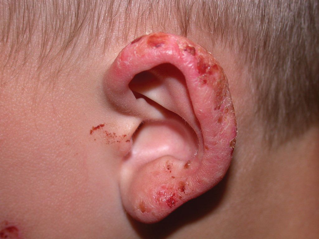
Juvenile spring eruption in a young boy
With Juvenile spring eruption (JSE), exposure to sunlight in early spring or summer results in a papulovesicular rash that usually resolves within 1 to 2 weeks.
Juvenile spring eruption | Image Credit: Author provided

Vignette
A 7-year-old boy developed an itchy, red, crusted rash on both ears within several hours after playing outdoors in the early spring sun. He was treated with emollients and a topical steroid with clearing over the next week and showed no signs of recurrence with reexposure to sunlight (Image). He had a similar eruption last spring. What’s the diagnosis?
Diagnosis Juvenile spring eruption
Introduction
Juvenile spring eruption (JSE) is a variant of polymorphous light eruption (PMLE), which is the most common form of light induced dermatosis. With JSE, exposure to sunlight in early spring or summer results in a papulovesicular rash that usually resolves within 1 to 2 weeks. The child typically presents with multiple red, edematous papules that may become vesicular and crusted on the helices of the ears. The lesions do not recur until the following spring. Another variant of JSE referred to as PMLE sine eruption presents with pruritus on sun-exposed skin without visible skin changes.1,2
Discussion
JSE is seen typically in boys and young men in early spring. While the exact etiology is not yet known, the immune-mediated nature of the reaction is widely supported. It is suggested that it occurs as a delayed hypersensitivity reaction to an endogenous antigen expressed after exposure to sunlight or artificial sources of radiation.1,3,4
Classic PMLE can occur over the face, the V area of the chest, the back of the neck, and the dorsolateral aspects of the forearms. The lesions may include grouped papules, eczematous plaques, vesicles, and plaques.2
Recurrent, unprotected sun exposure may result in recurrent lesions in cases of PMLE, which often settles down later in the summer. Polymorphous light eruption is also known as polymorphic light eruption, prurigo aestivalis, dermatographia photogenica, and erythema perstans solare.1,2
Literature reveals that in children, JSE has a moderate impact on the quality of life. If necessary, pruritus can be controlled with a topical steroid and oral antihistamines. Intense symptoms particularly in school-age children may require a short course of oral steroid. Thus, physicians may keep in mind the psychological aspects of the condition in their management.5
Differential diagnosis
Photosensitivity associated with lupus erythematosus and photosensitivity dermatitis are potential differentials. The lesions may appear similar but tend to be more persistent.1
Solar urticaria also manifests during or shortly after sun exposure. However, unlike JSE, it resolves within an hour or so of covering up from the source of light.1
Treatment
Management of JSE includes both preventive and curative measures.
Preventive
Fortunately, JSE is self-limited. However, the most important preventive measures include educating parents about the harmful effects of direct exposure to sunlight and UV rays, covering as much skin as possible, wearing full-sleeved clothing, and using broad-spectrum, water-resistant sunscreen with sun protection factor 50+ on exposed parts of the body including the face, ears, neck, and palms.1,6
Prior to exposure, short courses of hydroxychloroquine and antioxidants such as polypodium leucotomos extract, lycopene, beta-carotene, nicotinamide, and astaxanthin have some evidence of preventive value.1
For classic PMLE, phototherapy in late winter or early spring can reduce the incidence and severity of lesions. Two to 3 treatments per week for 4 to 6 weeks each year has shown to have prophylactic effect.1,6
Curative
- The first choice is topical steroids, which show reduction of lesions and itching due to their anti-inflammatory properties.
- Topical antioxidants like alphaglycosylrutin, ferulic acid, and tocopherol acetate were found to be useful in PMLE. These protect against the inflammatory reactions that are most likely to be mediated by the generation of free radicals in the skin.2
- Photochemotherapy also helps reduce the lesions through a phenomenon known as “hardening.”2
- Systemic corticosteroids and immunosuppressives are indicated in severe cases of classic PMLE.2,6
Our patient
The lesions subsided within a few days of application of topical hydrocortisone cream. The parent was advised of preventive measures and counseled because JSE resolves within a few weeks and does not recur until the following spring. Moreover, in most children recurrent episodes stop by puberty.
BIO
Aamir Anees is a medical student at the Kanachur Institute of Medical Sciences in Karnataka, India.
To read more from the April, 2023, issue of Contemporary Pediatrics®, click here.
References
1. Oakley AM, Ramsey ML. Polymorphic Light Eruption. StatPearls Publishing; 2022
2. Karthikeyan K, Aishwarya M. Polymorphous light eruption- an Indian scenario. Indian Dermatol Online J. 2021;12(2):211-219. doi:10.4103/idoj.IDOJ_434_20
3. Gruber-Wackernagel A, Byrne SN, Wolf P. Polymorphous light eruption: clinic aspects and pathogenesis. Dermatol Clin. 2014;32(3):315-viii. doi:10.1016/j.det.2014.03.012
4. Lava SA, Simonetti GD, Ragazzi M, Guarino Gubler S, Bianchetti MG. Juvenile spring eruption: an outbreak report and systematic review of the literature. Br J Dermatol. 2013;168(5):1066-1072. doi:10.1111/bjd.12197
5. Rizwan M, Haylett AK, Richards HL, Ling TC, Rhodes LE. Impact of photosensitivity disorders on the life quality of children. Photodermatol Photoimmunol Photomed. 2012;28(6):290-292. doi:10.1111/j.1600-0781.2012.00691.x
6. Ling TC, Gibbs NK, Rhodes LE. Treatment of polymorphic light eruption. Photodermatol Photoimmunol Photomed. 2003;19(5):217-227. doi:10.1034/j.1600-0781.2003.00048.x

Recognize & Refer: Hemangiomas in pediatrics
July 17th 2019Contemporary Pediatrics sits down exclusively with Sheila Fallon Friedlander, MD, a professor dermatology and pediatrics, to discuss the one key condition for which she believes community pediatricians should be especially aware-hemangiomas.
Itchy skin associated with sleep problems in infants
September 27th 2024A recent study presented at the American Academy of Pediatrics 2024 National Conference & Exhibition, sheds light on the connection between skin conditions and sleep disturbances in infants and toddlers, highlighting itchy skin as a significant factor, even in the absence of atopic