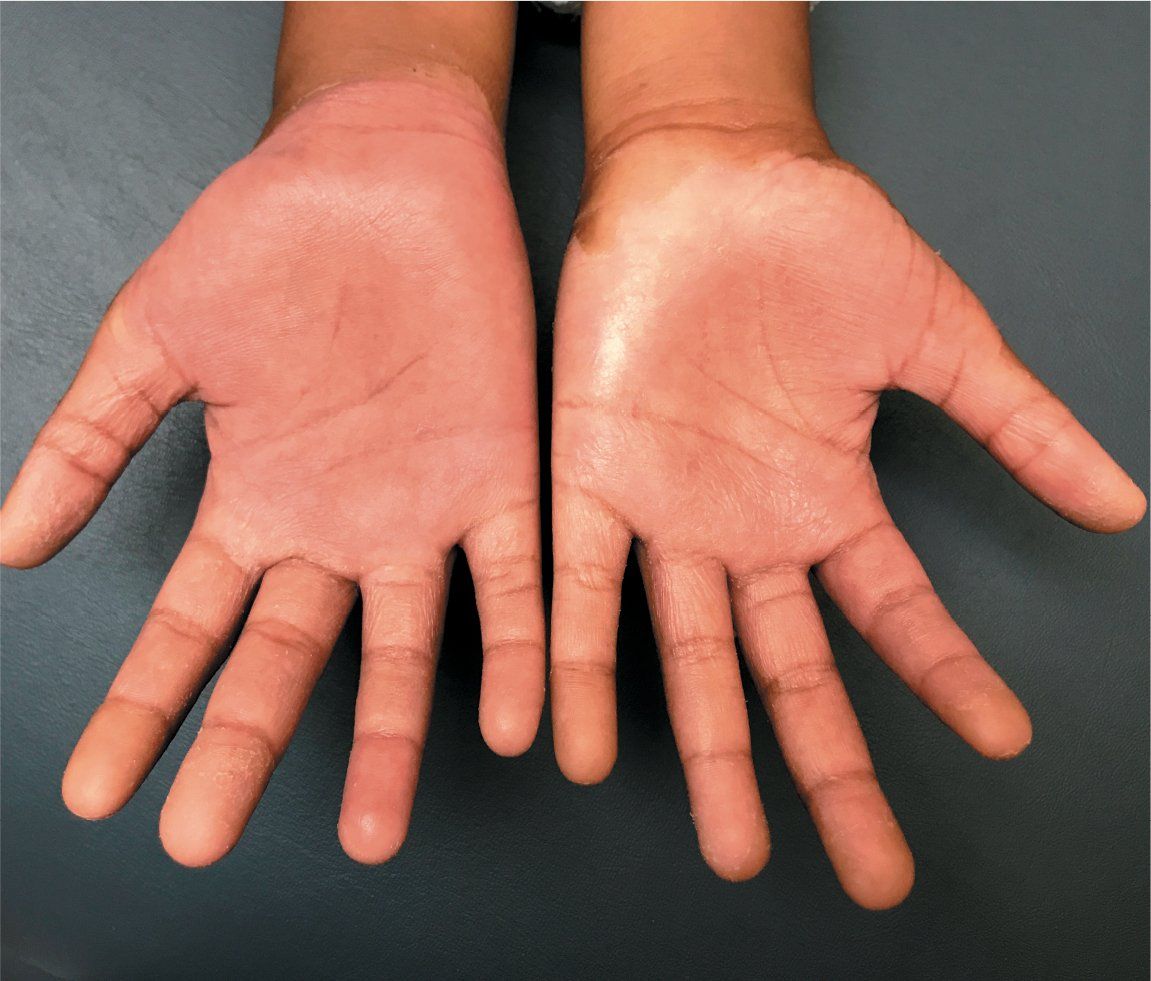
Painful, tense acral bullae in a 12-year-old girl
A healthy 12-year-old girl presents to the clinic with 2 days of low-grade fever and enlarging, painful, tense bullae on both hands. She had recently been diagnosed with streptococcal pharyngitis and was being treated with oral cefixime.
Patient at follow-up appointment showing remarkable recovery

The case
A healthy 12-year-old girl presents to the clinic with 2 days of low-grade fever and enlarging, painful, tense bullae on both hands. She had recently been diagnosed with streptococcal pharyngitis and was being treated with oral cefixime.
Diagnosis: Blistering distal dactylitis
Blistering distal dactylitis (BDD) is a rare, localized skin infection first described by Hays and Mullard in 1972 with poorly understood pathogenesis, most commonly caused by Group A beta-hemolytic Streptococcus and less commonly methicillin-sensitive Staphylococcus aureus or methicillin-resistant S aureus (MRSA).1-4 It classically targets children aged 2 to 16 years, is rarely seen in adults or infants, and spares the elderly.3-8
Clinical findings
Blistering distal dactylitis presents as tense, seropurulent, fluid-filled bullae on the volar fat pad of the distal phalanx or, rarely, proximal or lateral nail folds.2 One case of recurrent BDD was reported on the right great toenail associated with an infection caused by an ingrown toenail9 but, to the authors’ knowledge, no cases of BDD have been reported on the proximal or middle phalanx or so extensively on the dorsal hand, as in this case.
Diagnosis/Management
The initial clinical diagnosis of BDD is best confirmed by bacterial culture. The classic appearance of BDD is characterized as multiple superficial, tense, acral oval bullae 10 mm to 30 mm in diameter with an erythematous base.4 A limited set of tests can support the clinical diagnosis such as C-reactive protein (CRP), erythrocyte sedimentation rate (ESR), and white blood cell (WBC) count to suggest infectious etiology.
Differential diagnosis for BDD includes contact dermatitis, bullous impetigo, fixed drug eruption, and insect bites. Contact dermatitis also can cause bullous eruptions, although not often as proliferative as the bullae seen in this patient, and does not correlate with leukocytosis or fever. Fixed drug eruptions or unusual reactions to insect bites can present as annular or bullous lesions but are generally not as edematous. Bullous impetigo associated with staphylococcal infections can present similarly, but vesicles and bullae are usually much smaller and bacterial culture is needed to confirm clinical suspicions.
Therapy involves treating the underlying infection with systemic and topical antibiotics as well as incision and drainage of bullae.8
Patient outcome
The girl’s parents denied any new exposures to chemicals, lotions, or detergents and also denied taking any medications other than cefixime or having a family history of similar rashes.
She was started empirically on intravenous ampicillin for streptococcal and staphylococcal coverage, clindamycin for empiric MRSA coverage, and mupirocin 2% ointment to open lesions. The bullae were sterilely drained and fluid was sent for culture, which revealed Group A beta-hemolytic Streptococcus. Other significant lab values included a leukocytosis of 18,300 WBC/mm3 (neutrophil predominant); elevated CRP of 1.87 mg/dL; and elevated ESR of 26 mm/hr. After drainage of the bullae and completion of a 10-day course of oral cephalexin, the patient’s bullae completely resolved.
The importance of this case as a teaching tool is due to its exuberant presentation of an otherwise classic pediatric skin infection.
References:
1. Hays GC, Mullard JA. Can nasal bacterial flora be predicted from clinical findings? Pediatrics. 1972;49(4):596-599.
2. Rich P. Overview of nail disorders-blistering distal dactylitis. UpToDate. Available at: https://www.uptodate.com/contents/overview-ofnaildisorderssearch=%20blistering%20distal%20dactylitis§ionRank=1&usage_type=default&anchor=H57682918&source=machineLearning&selectedTitle=1~1&display_rank=1#H57682918. Updated August 29, 2017. Accessed February 12, 2019.
3. Kollipara R, Downing C, Lee M, Guidry J, Robare-Stout S, Tyring S. Blistering distal dactylitis in an adult. J Cutan Med Surg. 2015;19(4):397-399.
4. Scheinfeld NS. Is blistering distal dactylitis a variant of bullous impetigo? Clin Exp Dermatol. 2007;32(3):314-316.
5. Parras F, Ezpeleta C, Romero J, Sendagorta E, Buzón L. Blistering distal dactylitis in an adult. Cutis. 1988;41(2):127-128.
6. Scheinfeld N. A review and report of blistering distal dactylitis due to Staphylococcus aureus in two HIV-positive men. Dermatol Online J. 2007;13(2):8.
7. Fretzayas A, Moustaki M, Tsagris V, Brozou T, NIcolaidou P. MRSA blistering distal dactylitis and review of reported cases. Pediatr Dermatol. 2011;28(4):433-435.
8. Lyon M, Doehring MC. Blistering distal dactylitis: a case series in children under nine months of age. J Emerg Med. 2004;26(4):421-423.
9. Telfer NR, Barth JH, Dawber RP. Recurrent blistering distal dactylitis of the great toe associated with an ingrowing toenail. Clin Exp Dermatol. 1989;14(5):380-381.

Recognize & Refer: Hemangiomas in pediatrics
July 17th 2019Contemporary Pediatrics sits down exclusively with Sheila Fallon Friedlander, MD, a professor dermatology and pediatrics, to discuss the one key condition for which she believes community pediatricians should be especially aware-hemangiomas.