Reticulated rash on boy’s lower extremities
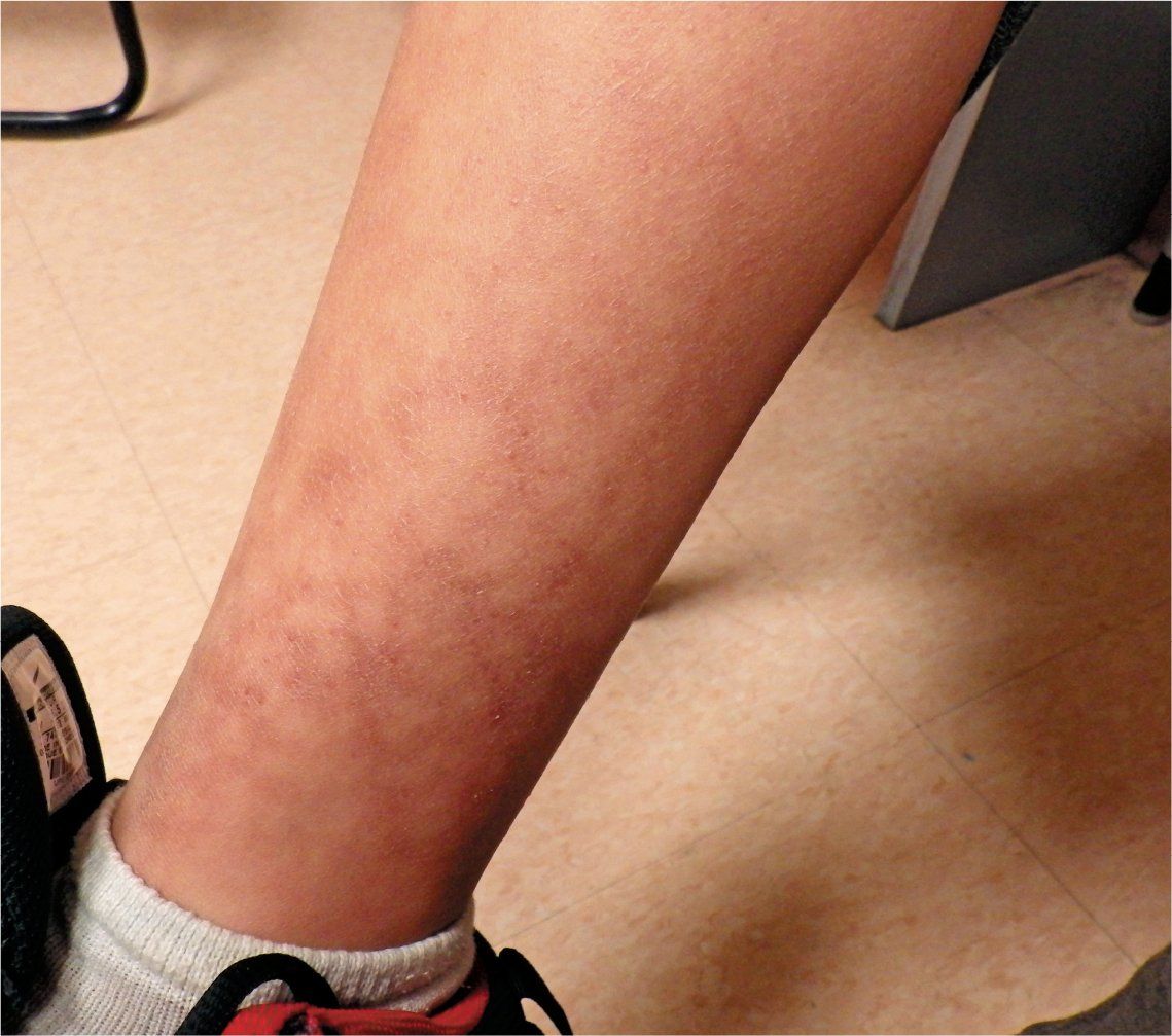
A healthy 11-year-old boy is brought to the office for evaluation of asymptomatic reticulated rash that started on his ankles 3 weeks ago and since has spread to his shins and the tops of both feet. He is on no medications and has had no history of trauma or recent illness. What's the diagnosis?
Patient presents with a persistent, nonblanching reticular rash on his lower extremities

A healthy 11-year-old boy is brought to the office for evaluation of asymptomatic reticulated rash that started on his ankles 3 weeks ago and since has spread to his shins and the tops of both feet. He is on no medications and has had no history of trauma or recent illness.
The patient was a well-developed, well-nourished child. Examination of the affected area showed a nonblanching, nontender, dark brown-to-purple reticular discoloration over both shins, ankles, and dorsa of feet. The remainder of the physical examination was normal.
Differential diagnosis
The differential diagnosis for reticular rash includes livedo reticularis, Henoch-Schonlein purpura (HSP), cutis marmorata telangiectatica congenital (CMTC), and erythema ab igne (EAI).
Livedo reticularis is a skin condition characterized by a mottled reticular pattern with purplish discoloration caused by impaired blood flow to the skin. It may be physiologic and reversible (cutis marmorata in infants exposed to cold) or pathologic and persistent1 secondary to vasculitis, systemic lupus erythematosus, drugs/toxin, cryoglobulinemia, antiphospholipid syndrome, and other connective tissue disorders. Many of these individuals with livedo reticularis also will have features that are suggestive of the underlying cause.
Henoch-Schonlein purpura is an immunoglobulin (Ig) A-mediated small vessel vasculitis resulting in a purplish, petechial, or purpuric rash on the dependent part of the body, namely the legs and buttocks. The face, arms, and torso also may be affected. It is most commonly seen in young children, generally between ages 2 to 8 years, and is often triggered by infections. Arthralgia, hematuria, abdominal pain, and scrotal edema may indicate involvement of other organ systems. Henoch-Schonlein purpura is a self-limited illness, but these patients may require long-term follow-up for the development of subtle renal complications.
Cutis marmorata telangiectatica congenita (CMTC) is a congenital venous anomaly characterized by persistent reticulated blanching erythema and subtle atrophy. Occasionally, ulcerations along the lines of atrophy may develop. It also can be associated with other anomalies such as limb asymmetry, port-wine stain, macrocephaly, and skeletal abnormalities such as clubfoot, hip dysplasia, and syndactyly.2 The vascular pattern is usually segmental and most likely represents a genetic mosaic condition.
Diagnosis: Erythema ab igne
Discussion
Erythema ab igne (which translates from Latin as “redness from fire”), also known as hot water bottle rash, toasted skin syndrome, ephelis ignealis, erythema caloric, or fire stains, is a skin condition characterized by localized areas of reticulated erythema and hyperpigmentation caused by chronic exposure to heat (infrared radiation) at a level lower than is required to cause a burn.3 Rarely, EAI may take on a bullous form.4 The most common reported causes are hot water bottles and electric heating pads, but any heat source can potentially cause EAI, including laptops, space heaters, and heated seats.
Initially, EAI starts out as a mild erythema over the exposed areas. With repeated exposure, a reticular, mottled, hyperpigmented, brown, blue, or purple rash develops.5 Erythema ab igne is typically a benign and self-limiting rash that resolves with discontinuation of the heat exposure. However, chronic exposure to heat can lead to more permanent color changes, dysplasia, and, in rare cases, carcinoma.6
Erythema ab igne is generally seen in individuals using heating devices to relieve local pain or cold. However, cases in which there was inadvertent exposure to heat, such as prolonged use of laptops placed over the thighs, and in professionals including jewelers or bakers who work with flames, also have been reported. On further inquiry, the patient revealed that he would spend the morning sitting in front of an electric base heater after he had showered to combat the New England winter.
Epidemiology/Pathophysiology
Erythema ab igne is uncommon, especially in countries such as the United States, where central heating is available. The use of open fires for warmth in underdeveloped countries is a known cause. It was also more common in populations whose work brought them into close contact with open flames or coal stoves. In developed countries, the condition is more likely to be seen in individuals who use local heating solutions such as electric heating pads or hot water bottles for pain relief. It occurs primarily in adults, with women being affected more than men. It is commonly seen on the lower back, legs, and abdomen, but can affect any body surface. There is an emerging trend of EAI in children using laptops on their thighs for extended periods of time.
The pathogenesis of EAI remains poorly understood. It is widely accepted that repeated heat exposure induces injury to the superficial blood vessels with hemosiderin deposition in the skin resulting in the characteristic reticular pattern. Microscopically, vacuolar degeneration at the dermoepidermal junction, with necrotic keratinocytes and melanin deposition, are characteristic of EAI.6 Squamous cell carcinoma and Merkel cell carcinomas may rarely develop This is much more common in adults and was a common phenomenon in the southern United States when fireplaces were the primary source of heating during the short winter season.7,8
Management
There is no definitive therapy for EAI. Elimination of exposure to heat may reverse the erythema and hyperpigmentation, especially if early in the disease process.3 More advanced cases require topical steroids or tretinoin to reduce the discoloration. Also, 5-fluorouracil has been shown to help clear atypical cells.9 Mesoglycan with topical flavonoids has shown promise in reducing the discoloration.10 Treatment with lasers is another option to reduce the pigmentary changes. More important is the long-term monitoring and follow-up for the development of cutaneous malignancies.
Patient outcome
The patient was advised to avoid sitting by the heater for prolonged periods of time. His lesions resolved over a period of several weeks without other intervention.
References:
1. Sajjan VV, Lunge S, Swamy MB, Pandit AM. Livedo reticularis: a review of the literature. Indian Dermatol Online J. 2015;6(5):315-321.
2. Levy R, Lam JM. Cutis marmorata telangiectatica congenita: a mimicker of a common disorder. CMAJ. 2011;183(4):e249-e251.
3. Baruchin AM. Erythema ab igne-a neglected entity? Burns. 1994;20(5):460-462.
4. Kokturk A, Kaya TI, Baz K, Yazici AC, Apa DD, Ikizoglu G. Bullous erythema ab igne. Dermatol Online J. 2003;9(3):18.
5. Dvoretzky I, Silverman NR. Reticular erythema of the lower back. Arch Dermatol. 1991;127(3):407-408.
6. Hurwitz RM, Tisserand ME. Erythema ab igne. Arch Dermatol. 1987;123(1):21-23.
7. LoPiccolo M, Crestanello J, Yoo SS, Sciubba J, Fernández C, Tausk FA. Facial erythema ab igne of rapid onset. Oral Surg Oral Med Oral Pathol Oral Radiol Endod. 2008;105(5):e38-e40.
8. Rudolph CM, Soyer HP, Wolf P, Kerl H. Squamous epithelial carcinoma in erythema ab igne [article in German]. Hautarzt. 2000;51(4):260-263.
9. Sahl WJ Jr, Taira JW. Erythema ab igne: treatment with 5-florouracil cream. J Am Acad Dermatol. 1992;27(1):109-110.
10. Gianfaldoni S, Gianfaldoni R, Tchernev G, Lotti J, Wollina U, Lotti T. Erythema ab igne successfully treated with mesoglycan and bioflavonoids: a case report. Open Access Mace J Med Sci. 2017;5(4);432-435.

Recognize & Refer: Hemangiomas in pediatrics
July 17th 2019Contemporary Pediatrics sits down exclusively with Sheila Fallon Friedlander, MD, a professor dermatology and pediatrics, to discuss the one key condition for which she believes community pediatricians should be especially aware-hemangiomas.