Toddler With Decreased Appetite and Activity
An 18-month-old white boy is brought to his well-care visit by his parents, who are concerned that for the past month he has been less social and active. He has appeared weak and has refused to walk or play with his siblings. He has also had a decreased appetite and has lost about 2 kg. He has vomited several times but with no bile or blood.
An 18-month-old white boy is brought to his well-care visit by his parents, who are concerned that for the past month he has been less social and active. He has appeared weak and has refused to walk or play with his siblings. He has also had a decreased appetite and has lost about 2 kg. He has vomited several times but with no bile or blood.
HISTORY
The child’s last well care visit was at age 6 months. He has had no recent illnesses, fever, diarrhea, or rash. He has a history of pica; recently, he was found ingesting fibers from the living room carpet. There is no family history of neurological, GI, rheumatological, or hematological disease. His immunizations are up-to-date. His diet consists of whole milk (six 4-oz bottles per day) and jarred baby foods. Developmental milestones have been appropriate. He lives with his mother and father and 2 brothers who are healthy.
PHYSICAL EXAMINATION
The child looks tired and pale. His weight is 8.9 kg (less than the 3rd percentile); height, 79 cm (10th percentile); and head circumference, 48.5 cm (50th percentile). Temperature is 36.9°C (98.4°F); heart rate, 160 beats per minute; respiration rate, 28 breaths per minute; blood pressure, 85/49 mm Hg; and oxygen saturation, 100% on room air. In addition to tachycardia, a 2/6 systolic ejection murmur is noted. Heart sounds are normal. He is mesocephalic and has clear sclerae, normal oral mucosa, and a supple neck. Chest and lungs are clear. Abdomen is soft with no organomegaly or masses. The skin on his palms and soles is also pale. He has no bruising or petechiae. He refuses to walk, but neurological findings are nonfocal.
LABORATORY RESULTS
Hemoglobin level is 2.7 g/dL. The white blood cell count is 8300/μL, with 13% segmented neutrophils and 79% lymphocytes; the platelet count is 597,000/μL. Mean corpuscular volume is 46 fL, red cell distribution width is 37%, and reticulocyte count is 2.2%. Results of a basic metabolic panel are normal. Transaminase, alkaline phosphatase, and bilirubin levels are also normal.
Which of the following would be the logical next step?A. Rule out an oncological process with a bone marrow biopsy.
B. Measure total iron and total iron-binding capacity, review the blood smear, and perform a Coombs test.
C. Obtain a better history related to possible ingestion as a cause of this severe anemia.
D. Refer to a hematologist.
(Answer and discussion on next page.)
The correct answer is B
About Morning Report:
When Chief Residents Share Their Challenging Cases, You Pass the Test
Send "Morning Report" submissions to:
linda.geisler@cmpmedica.com
Abnormally low hemoglobin can be the result of either a rapid breakdown of red blood cells (hemolytic anemia) or a failure of production in the bone marrow (iron deficiency anemia). The total serum iron level and total iron-binding capacity, a review of the blood smear, and results of a Coombs test can all help distinguish between these 2 causes. This child’s total serum iron level was low at 10 μg/dL (normal, 50 to 120 μg/dL), and the total iron-binding capacity was elevated at 496 μg/dL (normal, 250 to 400 μg/dL), because of insufficient iron levels in the body. The patient’s blood type was O positive; results of a Coombs test were negative; the bilirubin level was normal; and the blood smear showed severe microcytosis and hypochromia, but no basophilic stippling or fragmented cells. These findings made lead intoxication or hemolytic anemia unlikely. Because there was no hemolysis, bone marrow production problems were immediately considered. Results of hemoglobin electrophoresis were normal, and stools were hemoccult-negative. Iron deficiency anemia was the final diagnosis.
Hemoglobin electrophoresis is usually unnecessary when a newborn screen is available. Because this child was born in another state and was seen on a weekend, we were unable to access his birth records. In addition, the level of serum ferritin, an iron-storage protein, would most likely have been low, and the level of free erythrocyte protoporphyrin, a heme precursor, would have been elevated.
Hemoglobin levels are typically checked as part of a well-care visit between 9 and 12 months of age; however, this patient had not been seen since the age of 6 months, so this information was unavailable as well. Hemoglobin levels should also be routinely checked in older children (18 to 24 months), especially when the history suggests a high risk of iron deficiency anemia, and in children with special health needs.

It would have been reasonable to obtain a better history to ascertain the cause of the severe anemia (choice C) if lead intoxication was a concern; however, review of the blood smear would reveal basophilic stippling if the child had lead poisoning. Bone marrow biopsy (choice A) and referral to a hematologist (choice D) are a bit premature at this stage.
Clinical significance. Iron deficiency anemia is the most common nutritional deficiency in children; the peak incidence is between the ages of 6 months and 2 years. Infants are uniquely susceptible to this type of anemia because their dietary requirements for iron increase along with their rapid growth and expanding red blood cell mass. For this reason, iron supplementation is needed in exclusively breastfed infants by 4 months of age and in preterm infants soon after birth. Iron deficiency anemia can have significant clinical consequences. Poor oxygen delivery to organs can cause end-organ damage, heart failure, impairment of psychomotor and cognitive function, and even decreased leukocyte and lymphocyte function.

See erratum regarding Practice Pearl 2 below.
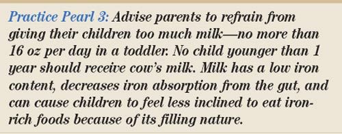
The correct amount of milk. In general, the appropriate amount of whole milk or 2% milk for toddlers is 16 oz per day. Parents should avoid giving their children too much milk because of the overall lack of iron in cow’s milk and because milk decreases iron absorption from the gut. In addition, children who drink a lot of milk feel full and as a result may be less inclined to eat ironrich foods. Parents are advised to wait until after the child is 12 months old to start whole milk, because of the occult GI bleeding that can occur from a cow’s milk allergy or enteropathy. However, cow’s milk allergy can cause occult GI bleeding even after 12 months of age.

Outcome of this case. The child was admitted and underwent emergent transfusion of packed red blood cells. Elemental iron therapy was started, and a nutritional consultation to reinforce an appropriate diet was recommended. After treatment, the child’s energy level returned to normal. Within 2 weeks, his hemoglobin level was normal for his age.
ONLINE EXCLUSIVE ...
Transfusion dosing. Children with symptomatic anemia (tachycardia, generalized weakness, and pica) require emergent transfusion of packed red blood cells. In a child with severe anemia, the transfusion should be made in aliquots over time to prevent overcompensation, which could place the child at risk for heart failure. If the patient’s hemoglobin level is greater than 5 g/dL, then the suggested dosing is 5 mL/kg over 3 to 4 hours times 3 aliquots. If the hemoglobin level is less than 5 g/dL, then the dosage is the value of the hemoglobin level (in mL per kg) (eg, if a child’s hemoglobin level was 3 g/dL and his weight was 3 kg, he would receive 9 mL of blood over 4 hours). Monitoring the patient’s cardiac function for signs of hypervolemia is essential; often patients need diuretics to help with volume overload.

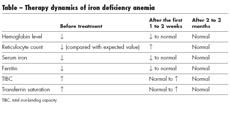
Iron therapy. Children with iron deficiency can be given 3 to 6 mg/kg of elemental iron per day. This should be taken with some source of vitamin C, generally orange juice, which serves as a cotransporter. The daily dose of iron does not need to be divided into 3 smaller doses, each given at a separate time of day; it is still very effective when given once daily. Laboratory values that can be used to monitor the child’s response to therapy are included in the Table.
Other conditions associated with anemia. Fanconi syndrome and Diamond-Blackfan syndrome occur in the first year of life; affected children have short stature and thumb anomalies. These syndromes are unlikely in children with normal developmental milestones and normal height and head circumference parameters.
Another, more common entity is transient erythroblastopenia of childhood. This usually develops after the first year of life. Affected children have a normocytic, normochromic anemia with no physical stigmata. As the name implies, this anemia is short-lived and has an excellent prognosis for recovery.
References:
FOR MORE INFORMATION:
• Beard JL. Why iron deficiency is important in infant development.
J Nutr.
2008;138:2534-2536.
• Flygare J, Karlsson S. Diamond-Blackfan anemia: erythropoiesis lost in translation.
Blood.
2007;109:3152-3154.
• Kazal LA Jr. Prevention of iron deficiency in infants and toddlers.
Am Fam Physician.
2002;66:1217-1224.
• Kohli-Kumar M. Screening for anemia in children: AAP recommendations-a critique.
Pediatrics.
2001;108:E56.
• Shaw J, Meeder R. Transient erythroblastopenia of childhood in siblings: case report and review of the literature.
J Pediatr Hematol Oncol
. 2007;29:659-660.
• Zlotkin S, Arthur P, Antwi KY, Yeung G. Randomized, controlled trial of single versus 3-times-daily ferrous sulfate drops for treatment of anemia.
Pediatrics.
2001;108:613-616.
Recognize & Refer: Hemangiomas in pediatrics
July 17th 2019Contemporary Pediatrics sits down exclusively with Sheila Fallon Friedlander, MD, a professor dermatology and pediatrics, to discuss the one key condition for which she believes community pediatricians should be especially aware-hemangiomas.