Ultrasonography: Expanding bedside applications in the pediatric emergency department setting
Bedside emergency ultrasonography has become a valuable tool for emergency physicians.
Remember those days searching in vain for vascular access in the child with sickle-cell disease who always was a difficult stick? Or the febrile toddler with a red foot that you clinically couldn't decide was an abscess, cellulitis, or an infection caused by a possible subcutaneous foreign body? Or the adolescent girl with a recent positive pregnancy test who was complaining of abdominal pain and vaginal bleeding?
In a busy emergency department (ED), great importance is placed on improving workflow and patient care. Bedside emergency ultrasonography has become a valuable tool for the emergency physician for just these reasons. It is a safe, rapid, goal-directed examination used to answer a focused and important clinical question, often in a yes-or-no fashion. Although it has been well established in the adult emergency setting over the past 20 years, its use is just gaining momentum in the pediatric emergency setting.
Children are the ideal patients for ultrasonography because their smaller bodies allow better penetration of the ultrasound waves compared with adults. Growing concerns regarding the small but significant long-term risks of inducing a fatal cancer as a consequence of ionizing radiation from computed tomography (CT) also makes ultrasound an attractive first-line imaging modality.1
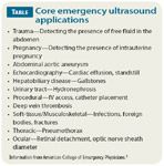
Emergency ultrasonography (EUS) has found its way into the pediatric ED largely because of the influence of our adult emergency medicine colleagues. The American College of Emergency Physicians (ACEP) has had an established policy statement on ultrasound in the ED since 2001 (updated in 2009) that describes the supporting literature, training pathways, credentialing, and quality management for EUS in the ED.2 These guidelines coincided with ultrasonography becoming an essential part of the emergency medicine residency curriculum and a core component in the practice of emergency medicine.

Diagnostic
TRAUMA
An adolescent male is brought into the ED by EMS after being thrown off his bike from a collision with a motor vehicle. He is dazed and tachycardic and has multiple abrasions, including on his abdomen. After the initial trauma resuscitation and stabilization, questions are asked: Is there an intra-abdominal solid organ injury, such as a lacerated spleen or liver? Will this patient need to go to the operating room if fluid resuscitation doesn't correct his tachycardia, and for what injury? Can you get these answers without leaving the bedside?
The Focused Assessment with Sonography for Trauma (FAST, or E-FAST when including lung views) examination has largely replaced previously more invasive and nonspecific investigations for free intraperitoneal blood to diagnose solid organ injury in the setting of blunt abdominal trauma. The examination looks at 4 sonographic "windows" where free, dependent blood can collect:
1. The right upper quadrant view looks for fluid within Morrison's pouch, a potential space situated between the liver and kidney.
2. The left upper quadrant view evaluates the space between the diaphragm and spleen.
3. The subxiphoid view looks for fluid within the pericardium.
4. The pelvic view looks for fluid posterior to the uterus in the pouch of Douglas for women or posterior to the bladder in men.
Although CT gives a more detailed picture of the abdominal anatomy, it requires that the trauma patient leave the controlled and monitored setting of an ED trauma bay, exposes the patient to ionizing radiation, and requires additional input from specialists who may not be immediately available. Blood from an injured organ such as the spleen, liver, or kidney settles into easily identifiable locations within the abdomen of a supine patient. This free fluid-whether blood, urine, or amniotic fluid-appears black or "hypoechoic" next to the relatively bright or "hyperechoic" surrounding solid organs. In the unstable trauma patient, a positive FAST identifies the source of blood loss and may help to direct further operative management.
In the setting of blunt abdominal trauma in an adult, a positive FAST and hypotension indicates the need for exploratory laparotomy. Whereas the use of the FAST examination as a screening tool for abdominal solid organ injury has been well established in the adult literature,3 pediatric blunt abdominal injuries are not as straightforward. Pediatric solid organ injuries often remain intracapsular, without free fluid in the abdomen. A study of stable pediatric trauma patients found that about half of those with an abdominal solid organ injury detected on CT had a negative FAST.4
A more recent meta-analysis including nearly 4,000 patients determined the sensitivity of FAST in children to be 80%, with a specificity of 96%, making it a useful tool in the detection of free fluid but not as a screen for abdominal injury.5 Perhaps this is why only 15% of pediatric stand-alone trauma centers use FAST, whereas 85% of centers that see both adult and pediatric patients routinely use FAST in the workup of blunt abdominal trauma.6
The question remains whether the injuries missed by FAST are clinically significant. The majority of solid organ injuries missed on FAST are low-grade lesions that do not require operative management. Further refinement on how to use FAST as a screening tool found that the sensitivity increased to 100% when including pertinent physical exam findings.7
A positive FAST in the hypotensive trauma patient identifies the source of blood loss and helps to aid the surgical management. In the hemodynamically stable patient, the FAST examination should be used in conjunction with the history, physical examination, and laboratory findings.
Trend talk: Increasing THC ingestions in pediatric emergency departments
October 4th 2024At the 2024 AAP National Conference, Steven Selbst, MD, highlighted the rise in accidental THC ingestions among children, urging pediatricians to counsel parents on the dangers of improperly stored THC gummies that resemble candy.
Study: Child heat-related illness ED visits up 170% in past decade
September 28th 2024“The significant rise in heat-related illnesses over the past decade underscores a growing public health concern that warrants further attention and action, said Taylor Merritt, MD, of the data presented at the AAP National Conference & Exhibition.