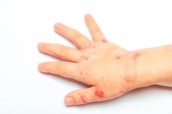
Bullous Impetigo in a Neonate
Multiple tender flaccid bullae (about 2 mm to 2 cm) were present on the perineum. There was no associated discharge. The ruptured blisters had marked erythema in the center and a scaly rim at the periphery. The Nikolsky sign could not be elicited. The infant was afebrile. The remainder of the examination findings were normal.
FigureSeven days after birth, a baby boy was brought for evaluation of a "diaper rash." The eruption began 2 days earlier as a few vesicles and progressed to large blisters. Some of the lesions had burst. There was no history of fever. The infant was born at term via normal vaginal delivery. He was breast-feeding adequately. The mother tested negative for group B
Streptococcus
infection. She had no history suggestive of mastitis. There was no family history of skin infection.
Multiple tender flaccid bullae (about 2 mm to 2 cm) were present on the perineum. There was no associated discharge. The ruptured blisters had marked erythema in the center and a scaly rim at the periphery. The Nikolsky sign could not be elicited. The infant was afebrile. The remainder of the examination findings were normal.
Bullous impetigo (BI) was diagnosed. This common, highly contagious, superficial blistering skin infection is now recognized as a localized form of staphylococcal scalded skin syndrome (SSSS), or Ritter disease.1,2 The Table lists the differences between the 2 conditions.
Table
BI is caused by coagulase-positive Staphylococcus aureus (most commonly phage type II) or, rarely, by group A ?-hemolytic Streptococcus, either alone or in combination with S aureus. Culture helps identify the cause, which in this patient was methicillin-sensitive S aureus. Methicillin-resistant S aureus may be isolated. Results of antistreptolysin O and streptozyme tests may be positive but are rarely performed.1,2
Humans are a natural reservoir of S aureus; up to 50% of the general population are nasal carriers of this bacteria.2 In neonates, S aureus is commonly found on the skin, around the eyes, umbilicus, perineum, wound sites, circumcision wounds, and umbilical stumps.2 In one literature review, S aureus was isolated in 72% of 2482 skin and soft tissue infections in children.3 Predisposing factors for staphylococcal infection include immunodeficiency, diabetes, chronic granulomatous disease, skin breakdown (surgery, insect bites, varicella, herpes simplex), burns, and foreign substances (sutures, shunts, intravascular catheters).1,2
BI begins as a single red macule or papule that quickly becomes a vesicle. The vesicle rapidly enlarges into flaccid bullae with sharp margins without surrounding erythema. The bullae may rupture, ooze, and leave yellow crusts. The rash spreads by autoinoculation. A pathognomonic finding is a collarette of scale around the blister roof at the periphery of ruptured lesions.1,2 Moist, intertriginous areas, such as the diaper area, axillae, and neck folds, are commonly affected. The lesions may be mistaken for child abuse (eg, localized disease may mimic cigarette burns and more extensive infection may resemble scald injuries).4 Systemic symptoms are uncommon. Most cases are self-limited and resolve without scarring within several weeks.
Medical treatment of neonates who have BI consists of parenteral antistaphylococcal antibiotics. Control of pain, temperature, and hydration is essential. Coverage for secondary Gram-negative infection is recommended in severe cases. Exfoliation usually continues for 24 to 48 hours after starting antibiotics.1,2
This patient responded well to treatment with intravenous clindamycin and was discharged after 72 hours.
References:
- Cole C, Gazewood J. Diagnosis and treatment of impetigo. Am Fam Physician. 2007;75:859-864.
- Ladhani S, Garbash M. Staphylococcal skin infection in children-rational drug therapy recommendations. Pediatr Drugs. 2005;7:77-102.
- Darmstadt GL. Oral antibiotic therapy for uncomplicated bacterial skin infections in children. Pediatr Infect Dis J. 1997;16:227-240.
- Koning S, Verhagan AP, van Suijlekom-Smit LW, et al. Interventions for impetigo. Cochrane Database Syst Rev. 2004;(2):CD003261.
- Sandhu K, Kanwar AJ. Generalized bullous impetigo in a neonate. Pediatr Dermatol. 2004;21:667-669.
- Stanley JR, Amagai M. Pemphigus, bullous impetigo and the staphylococcal scalded skin syndrome. N Engl J Med. 2006;355:1800-1810.
- Hanakawa Y, Schechter NM, Lin C, et al. Molecular mechanisms of blister formation in bullous impetigo and staphylococcal scalded skin syndrome. J Clin Invest. 2002;110:53-60.
Newsletter
Access practical, evidence-based guidance to support better care for our youngest patients. Join our email list for the latest clinical updates.


