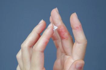
Newborn's rash linked to mother's antibodies
You are called from your office to the nursery to see a healthy newborn with a diffuse facial eruption. Her mother, who had a normal pregnancy, labor, and delivery, has a history of Sjogren's syndrome.
THE CASE
You are called from your office to the nursery to see a healthy newborn with a diffuse facial eruption. Her mother, who had a normal pregnancy, labor, and delivery, has a history of Sjögren's syndrome. Both mother and baby have SSA/Ro and SSB/La antibodies. Other laboratory studies, including a complete blood count and liver function tests, and a cardiac examination with an electrocardiogram were normal.
DIAGNOSIS: Neonatal lupus erythematosus
Cutaneous manifestations of NLE occur in 15% to 25% of cases.1 In most instances, a rash appears between 4 and 6 weeks but may present at birth or not until the infant reaches several months of age.1,4 The typical rash is characterized by 0.5-cm to 3-cm annular erythematous papules and plaques with atrophy, telangiectasias, dyspigmentation, and central scale.4
The rash frequently begins on the scalp and face, and periocular involvement is sometimes prominent, but it may spread to involve the neck, upper trunk, and upper extremities.1,4,5 It tends to be photosensitive but may appear in non-sun-exposed areas, including the palmar-plantar surfaces and diaper area. The rash is usually transient, lasting a mean duration of 15 to 17 weeks.1,6 Its disappearance coincides with the clearance of maternal IgG antibodies from the infant's circulation.4 However, in 10% to 20% of cases, subtle atrophy, telangiectasias, and pigmentary changes may remain.1,4
PATHOPHYSIOLOGY
More than 95% of cutaneous cases of NLE involve circulating SSA/Ro antibodies, but there are reported cases of only U1RNP antibodies being present.1 One study found that cutaneous NLE in an SSA/Ro antibody-exposed infant predicted a 6- to 10-fold risk of a subsequent pregnancy developing cardiac NLE.6 Other manifestations may be seen, including petechiae or purpura secondary to thrombocytopenia and jaundice secondary to hepatic involvement.7
Neonatal lupus erythematosus is the number one cause of congenital heart block.4 Cardiac complications of NLE are associated with significant morbidity and mortality.6 Nearly 67% of neonates with cardiac manifestations of NLE require pacemaker placement, and as many as 20% to 30% die.
Approximately 10% of cases of NLE are associated with liver and hematologic involvement.8 Hepatic involvement is typically characterized by an asymptomatic elevation in alanine aminotransferase and aspartate aminotransferase, but hyperbilirubinemia, hepatitis, hepatomegaly, splenomegaly, and even liver failure have been reported.1,8
Newsletter
Access practical, evidence-based guidance to support better care for our youngest patients. Join our email list for the latest clinical updates.


