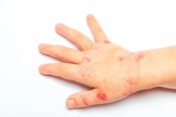
Unexplained Bruising in a Teenager: Abuse-or Mimic?
A 14-year-old boy presented to the abuse clinic at the request of child protective services. The boy’s gym teacher had noticed multiple large bruises on his back, arms, and legs. School officials had previous experience with the child’s “volatile” stepfather and thought him “capable of abuse.” There were also unofficial concerns of past domestic abuse. A 14-year-old boy presented to the abuse clinic at the request of child protective services. The boy’s gym teacher had noticed multiple large bruises on his back, arms, and legs. School officials had previous experience with the child’s “volatile” stepfather and thought him “capable of abuse.” There were also unofficial concerns of past domestic abuse.
A 14-year-old boy presented to the abuse clinic at the request of Child Protective Services (CPS). According to CPS, the boy’s gym teacher had noticed multiple large bruises on his back, arms, and legs. School officials had previous experience with the child’s “volatile” stepfather and thought him “capable of abuse.” There were also unofficial concerns of past domestic abuse per the CPS history.
The boy had been interviewed at school by teachers and CPS and had repeatedly denied any abuse. He alleged a very good relationship with his stepfather, and denied any form of trauma.
The boy’s family came to the abuse clinic without any pressure from CPS. They seemed more concerned about the child’s health and safety than any possible allegations being leveled at them. The parents provided a complete medical history while the boy was interviewed by a social worker. The family stated that the teen was very private and that they had not seen his bruises until the school called. The stepfather had been in the family for 12 years. While the boy was examined and the final aspects of the medical history were obtained, the family was interviewed by the social worker.
All the histories were consistent with one another. The history of volatility and domestic abuse was not verified by cursory investigation by law enforcement.
The medical history revealed that the boy had a viral infection in the past 2 to 3 weeks. The physical exam revealed 11 large bruises (Figure). There was no specific pattern or uniformity to the bruises. The boy was private and would only consent to our taking a picture of his arm. The abdominal exam was compatible with an enlarged spleen (3 cm below the costal margin).
After a meeting of the abuse clinic staff, CPS, and law enforcement before discharging the boy, all agreed on the diagnosis.
Was this physical abuse-or a mimic?
(Please
DIAGNOSIS: Immune thrombocytopenic purpura
The allegation of abuse was based on multiple incorrect assertions. A thorough medical history was adequate to establish a potential medical diagnosis. Law enforcement and CPS were invaluable in collecting a good social history.
The diagnosis was established with just a CBC. The hemoglobin was 14 g/dL; the WBC count was 4700/µL, with a normal differential; the platelet count was 8000/µL. A bone marrow aspirate revealed a cellular marrow, with a normal myeloid and erythroid maturation and increased megakaryocytes with increased young forms. The diagnosis was ITP.
The patient was treated with WinRho anti D and the platelet count rose to 70,000/µL by day 4.
It is not uncommon for patients with bruising to be referred to a pediatric hematologist-oncologist or child abuse clinic. Bruising in a child should always raise a the spectre of child abuse.1,2
In our patient’s case, the laboratory results were diagnostic of ITP. The most common cause is a platelet autoantibody produced after a viral infection.3,4 The antibodies coat the platelets, which are then taken up and destroyed by the spleen. The thrombocytopenia is thought to be secondary to platelet destruction. Recent work has shown that decreased platelet production also contributes to the thrombocytopenia. Other causes for ITP include an underlying autoimmune disorder, an immunologic dysfunction disease, drug-induced thrombocytopenia, and persistent viral infections.3,4,6
ITP post-viral infection is typically a self-limited condition in the pediatric population. Most children (70% to 80%) recover within 4 to 6 months. Chronic ITP (ie, thrombocytopenia that lasts longer than 1 year), occurs in 20% to 30% of the patient population and is more common in older children.3,4
The diagnosis of ITP is one of exclusion. Platelet antibody testing is not useful because of its low specificity.4 The affected child is clinically asymptomatic, but may have a history of recent viral illness. The physical examination is normal except for skin bruising, petechiae, and occasionally mild splenomegaly reflecting a recent viral illness.
Bone marrow aspiration is no longer recommended in the typical case of post-viral ITP.3,4
For affected patients who do not have severe mucosal bleeding, treatment includes watchful waiting. For those with more serious bleeding, corticosteroids, intravenous immunoglobulins, and Rh(D) immunoglobulin are the treatments of choice. In chronic ITP, androgens and chemotherapeutic agents have been tried as well as splenectomy. More recently, thrombopoietin receptor agents have been used to elevate platelet counts.3,4
A bone marrow aspiration was done in this boy’s case, to rule out underlying bone marrow dysfunction. It was also performed because of his age and because of the splenomegaly. The patient was Rh-positive and was given anti-Rh immunoglobulin (WinRho) in anticipation of a rapid rise in platelet count.3,4 In fact, he responded to a single dose of WinRho and maintained a safe platelet count.
References
1. Maguire S, Mann MK, Sibert J, et al. Are there patterns of bruising in childhood which are diagnostic or suggestive of abuse? A systematic review. Arch Dis Child. 2005;90:182-186.
2. Thomas AE. The bleeding child; is it NAI? Arch Dis Child. 2004;89:1163-1167.
3. Neunert C, Lim W, Crowther M, et al. The Americian Society of Hematology 2011 evidence-based practice guideline for immune thrombocytopenia. Blood. 2011;117:4190-4207.
4. Consolini DM. Thrombocytopenia in infants and children. Pediatr Rev. 2011;32:135-149.
5. Barsam SJ, Pasaila B, Forestier M. Platelet production and plate destruction: accessing mechanisms of treatment effect in immune thrombocytopenia. Blood. 2011;117:5729-5732.
6. Cines DB, Bussel JB, Liebman HA, et al. The ITP syndrome: pathogenic and clinical diversity. Blood. 2009;133:6511-6521.
Newsletter
Access practical, evidence-based guidance to support better care for our youngest patients. Join our email list for the latest clinical updates.


