In this podcast, Dr John Harrington of Eastern Virginia Medical School and Children’s Hospital of The King’s Daughters, and Dr Michael Paul, CEO and Rena Vanzo, Genetic Counselor of Lineagen-provider of a new integrated genetic testing and counseling service FirstStepDx-discuss the diagnosis of autism and genetic testing for autism.
John Harrington, MD
Advertisement
Articles by John Harrington, MD
A 3-year-old boy with chest pain and trouble breathing that had developed over the past 24 hours was brought to the emergency department. The parents reported that his most prominent symptom was a cough. The chest pain appeared to worsen with coughing. He had undergone open atrial septal defect repair about 3 weeks before presentation.
A 7-year-old boy with annular, asymptomatic, flesh-colored lesion onthe wrist that had developed slowly over the past month. The parents hadremoved the child from school because they were told that the lesion wasringworm. The lesion had failed to resolve after application of an antifungalcream for 10 days.
The cause was pilonidal sinus disease. The term pilonidal is derived from the Latin words "pilus," meaning "hair," and "nidus," meaning "nest." Pilonidal sinus disease is more common in males than in females and typically appears during adolescence. About 1% of all males and 0.1% of all females have an asymptomatic pilonidal sinus with potential for disease.1 The suspected overall incidence is about 1 in 5000. The disease seems to be most prevalent in those with stiff, dark or auburn hair, although it has been observed in all races.2
In this infant, microscopic examination of a potassium hydroxide preparation of scalp scrapings revealed Trichophyton tonsurans, which is the most common causative organism in North America.
IP is a rare X-linked dominant disorder. About 700 to 1000 cases have been reported worldwide (about 1 in 50,000 live births); white infants are most commonly affected. In a review of 653 patients, more than half had a family history of the condition.1 Our patient's mother was also affected. IP usually appears within the first 2 weeks of life. The severity and expression of the disorder are highly variable, even among patients within the same family.1,3 The condition is characterized by anomalies of the organs and tissues derived from the ectoderm and mesoderm and may affect the skin, hair, nails, teeth, eyes, and CNS1,2:
This baby was born with an ulnar supernumerary digit on the left hand. This common congenital anomaly can be an isolated malformation or associated with other syndromes at birth.1-4 Although the true prevalence is unknown (because most hospitals do not report cases), the condition appears to be more common among males and African American infants.1-3 In African American infants, postaxial polydactyly is generally the result of autosomal dominant transmission. In white infants, the condition is frequently syndromic and is linked with autosomal recessive transmission.1
One week after vaccination with diphtheria, tetanus, and acellular pertussis/inactivated poliovirus/hepatitis B, Haemophilus influenzae type b, pneumococcal conjugate, and rotavirus, this 2Z\x-month-old infant presented with a vesicular rash. No other children in the home had a rash. The infant's primary caregiver was the grandmother, who had shingles 2 weeks earlier.
The patient, a 13-year-old girl, was concerned about the development of a very itchy, painful, papular rash on her hands and feet. She had been previously well and had no history of illness other than a minor upper respiratory tract infection 2 weeks earlier. The distribution of lesions and the severe pruritus initially suggested scabies, which was treated with 5% permethrin cream. The rash did not improve with applications of this medication, however, and the patient returned the following day for care. She had no oral lesions but complained of mildly painful, nonswollen joints.
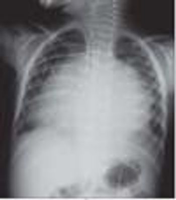
The patient is an 8-year-old girl with a history of asthma and developmental delay. She complained of hip pain, and her pediatrician referred her to a pediatric orthopedist for consultation. Hip x-ray films were ordered; they revealed 3 round beads in the child's appendix.
The lesions in this newborn's mouth are Bohn nodules. These mucous gland cysts manifest as firm, small (less than 3 mm) grayish white nodules--usually in the mouth on the buccal or lingual aspect of the alveolar ridges. They occasionally occur on the palate, and may be confused with large Epstein pearls.
Advertisement
Latest Updated Articles
 Making the Rounds
April 1st 2006
- Postaxial Polydactyly
November 1st 2007
- Photoclinic: Swallowed Beads
February 1st 2006
- Varicella
October 1st 2007
- Photoclinic: Bohn Nodules
October 1st 2005
- Sacrococcygeal Pilonidal Sinus
October 1st 2008
Advertisement
Advertisement
Trending on Contemporary Pediatrics
1
FAQ: Which children with early puberty actually need testing and treatment?
2
ACIP dropped MMRV coverage. New data show it was a lifeline for VFC-eligible kids
3
FDA grants tegacorat orphan, rare pediatric disease status for DMD
4
Douglas Mack, MD, shares details on the ongoing THRIVE study of epicutaneous immunotherapy for peanut allergy
5