- July 2021
- Volume 38
- Issue 7
A case of lime disease
A 17-year-old boy presents with linear brown spots on his abdomen and chest that developed when he was on a beach vacation with his family the week prior. What's the diagnosis?
The case
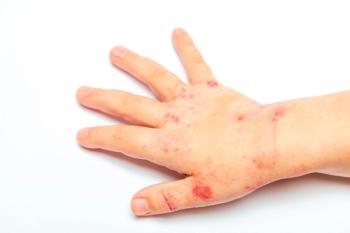
You are asked to evaluate a 17-year-old boy who developed linear brown spots on his abdomen and chest while on a beach vacation with his family the week prior. The patient first noticed the lesions a few days after he made a freshly squeezed limeade and spent the day at the beach. On examination, he has hyperpigmented macules of varying sizes located on the torso. Upon closer inspection, the lesions appear to have bizarre shapes consistent with streaks and drips. The lesions are asymptomatic (Figure).
Diagnosis: Lime disease/phytodermatitis
Discussion
Phytophotodermatitis is a cutaneous reaction caused by phototoxic compounds, present in certain plants, and subsequent sunlight exposure.1 These phototoxic compounds, known as furocoumarins, are produced by many common plants including lime, lemon, grapefruit, celery, carrot, parsnip, fig, celery, and dill.2,3 After the affected skin comes in contact with substances in these plants and is exposed to UV-A (280-315 nm) light, furocoumarins cause cellular damage and injury to the epidermis typical of sunburn.4 Signs of inflammation such as erythema, edema, and bullae typically form within 24 hours.4 Lesions appear only on sun exposed areas of the body that come in direct contact with the plant substances. They may take on bizarre forms, depending on how the phototoxic compounds come into contact with the skin. Lesions heal in 1 to 2 weeks and are replaced by hyperpigmentation, which may take several weeks or months to resolve.4 Short sunlight exposure may result in subtle asymptomatic erythema that goes unnoticed until the hyperpigmentation develops.
Differential diagnosis
The differential diagnosis for phytophotodermatitis includes contact dermatitis, bruising, and cellulitis. Contact dermatitis may be caused by irritants or external allergens. Irritant contact dermatitis (ICD) is more common and is nonimmune mediated. Anyone can develop ICD with sufficient exposure to a substance such as an acid, alkali, or oil. Previous contact with the irritant is not necessary, unlike in allergic contact dermatitis (ACD), which requires prior sensitization. ACD is a type IV hypersensitivity reaction. Common contact allergens include metals such as nickel, fragrances, preservatives, neomycin, rubber chemicals, and para-tertiary-butylphenol-formaldehyde resin.5 Unlike phytophotodermatitis lesions. which are often asymptomatic or minimally painful, the predominant symptom in contact dermatitis is pruritus. Bruising may be seen secondary to minor accidents and also to nonaccidental trauma. Phytophotodermatitis may be mistaken for child abuse, especially when it presents as well-demarcated “hand-shaped” marks and burnlike lesions.6 Unlike the lesions of phytophotodermatitis, bruises are not typically uniform in color. and they change hues as hemoglobin breaks down.7
Cellulitis is an infection of the dermis and subcutaneous tissue.8 Pain tends to be significant and accompanied by other classic signs of inflammation including redness, warmth, and swelling. In addition, the borders of cellulitis are usually poorly demarcated. The borders of phytophotodermatitis are well demarcated and limited to areas of phototoxic compound and sun exposure.
Treatment
Phytophotodermatitis is a self-limited condition and usually does not require treatment. Immediate treatment may involve removal of the offending plant substance and supportive care. Topical corticosteroids and cold compresses may alleviate pain.6
Patient outcome
Phytophotodermatitis was diagnosed clinically and treatment was not required. The teen was counseled on how to avoid future episodes including washing hands with soap and water after coming into contact with plants known to cause phytophotodermatitis. The importance of sun protection including the use of broad-spectrum sunscreen, wearing sun-protective clothing, and avoiding the sun during peak hours was also reviewed.
References
- Son J‐H, Jin H, You H‐S, et al. Five cases of phytophotodermatitis caused by fig leaves and relevant literature review. Ann Dermatol. 2017;29(1):86‐90. doi:10.5021/ad.2017.29.1.86
- Janda PH, Bhambri S, Del Rosso JQ, Mobini N. Phytophotodermatitis: case report and review of the literature. Cosmetic Dermatology. 2008;21(2):99-103.
- Pfurtscheller K, Trop M. Phototoxic plant burns: report of a case and review of topical wound treatment in children. Pediatr Dermatol. 2014;31(6):156-159. doi:10.1111/pde.12396
- Carlsen K, Weismann K. Phytophotodermatitis in 19 children admitted to hospital and their differential diagnoses: child abuse and herpes simplex virus infection. J Am Acad Dermatol. 2007;57(suppl 5):88-91. doi:10.1016/j.jaad.2006.08.034
- de Waard-van der Spek FB, Andersen KE, Darsow U, et al. Allergic contact dermatitis in children: which factors are relevant? (review of the literature). Pediatr Allergy Immunol. 2013;24(4):321-329. doi:10.1111/pai.12043
- Goskowicz MO, Friedlander SF, Eichenifield LF. Endemic “lime” disease: phytophotodermatitis in San Diego County. Pediatrics. 1994;93(5):828-830.
- Mathews MR, VanderVelde JC, Caruso DM, Foster KN. Lemons in the Arizona sunshine: the effects of furocoumarins leading to phytophotodermatitis and burn-like injuries. Wounds Research. 2017;29(12):118–124.
- Raff AB, Kroshinsky D. Cellulitis: a review. JAMA. 2016;316(3):325-337. doi:10.1001/jama.2016.8825
Articles in this issue
over 4 years ago
Allergies, asthma, and climate changeover 4 years ago
Gastrointestinal disorders: Red flags and best treatmentsover 4 years ago
A newborn with fever and tremorsover 4 years ago
Keeping your child safe from COVID-19 this summerover 4 years ago
Adjunctive medications for epilepsyover 4 years ago
Helping parents cope with adolescent behaviorover 4 years ago
Good quality of life may help overcome food allergyover 4 years ago
Participation in after-school activities decreases ADHD severityNewsletter
Access practical, evidence-based guidance to support better care for our youngest patients. Join our email list for the latest clinical updates.


