- October 2022
Case of inflammatory acne or something else?
This case study analyzes variants of majocchi granuloma and potential treatment.
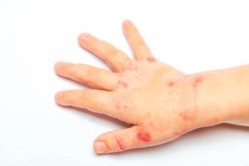
A healthy 12-year-old girl presented with an inflammatory acneiform eruption symmetrically distributed across her face with involvement of the eyelids and eyelid margins with styelike lesions. The patient reported intense itch and was subsequently treated with topical steroids, demonstrating a dramatic improvement only to recur as soon as she stopped application. A skin biopsy showed a granulomatous process with hyphae.
Introduction
Majocchi granuloma (MG) was discovered by Domenico Majocchi in 1883 as a subcutaneous or an intracutaneous inflammatory lesion resulting from a local invasion by the dermatophyte Trichophyton tonsurans. MG is a rare clinical form of tinea corporis infection.1 It is also known as Majocchi trichophytic granuloma, dermato-phytic granuloma, or nodular gran-ulomatous perifolliculitis.2
The 2 main clinical variants of MG are a papulopustular perifollicular form that develops after trauma in healthy individuals and a nodular form with indurated plaques and erythematous subcutaneous nodules presenting in immunocompromised patients.
The most common cause of MG is Trichophyton rubrum, followed by Trichophyton mentagro-phytes, Trichophyton violaceum, and T tonsurans.3
Discussion
MG is a global disease because causative organisms are present worldwide. Because the multiple erythematous nodules with ulceration and crusts seen on the face of the patient may have been perceived as psoriasis or another primary inflammatory noninfectious disorder, topical corticosteroids were prescribed. Dermatophytes usually cause superficial infection and do not invade beyond the reticular dermis. However, any mechanical trauma to the skin or immunocompromised state allows these fungi to penetrate beyond and cause deep infections. The species is keratinophilic and keratolytic, causing deeper invasion.4
Diagnosis of MG is confirmed through a potassium hydroxide mount and/or histopathological studies. Fungal hyphae are seen with branching and will reveal the causative organism. The specific species can be determined by fugal culture.
Differential diagnosis in healthy patients
Pustular psoriasis
Pustular psoriasis also presents as pustules with an erythematous background. However, with the 2 clinical subtypes—palmoplantar pustular psoriasis and generalized pustular psoriasis—the face is not usually affected. Moreover, the application of topical steroids would have cleared the lesions with no recurrence immediately after stopping the drug,5 which was not seen in our patient.
Acne vulgaris
Acne vulgaris may sometimes present as erythematous nodules with hyperpigmentation; however, the lesions in our patient were more severely inflammatory. Further, there were no comedones or cysts6 in our patient.
Granulomatous rosacea
Later stages of granulomatous rosacea may seem similar to the nodular lesions as seen in the patient; however, telangiectasias and permanent flushing are absent.7
Bacterial folliculitis
Bacterial folliculitis or infection of the hair follicles is commonly seen post shaving or use of unclean razors. However, histopathology would reveal bacterial organisms if it were bacterial folliculitis.
Treatment
With excellent treatment outcome and clearing of lesions, terbinafine is the preferred drug. It has both fungistatic and fungicidal action, with high efficacy and low drug interactions. Treatment should be continued until the lesions are cleared. Other options include itraconazole, voriconazole, and griseofulvin.8
Resolution
After reviewing the histopathological reports and analysis of the hyphae seen in microscopy, we were able to confirm the fungal etiology of the lesions. Oral terbinafine therapy was initiated for 3 weeks with clearing of the lesions.
Reference
1. Ilkit M, Durdu M, Karakaş M. Majocchi’s granuloma: a symptom complex caused by fungal pathogens. Med Mycol. 2012;50(5):449-457. doi:10.3109/13693786.2012.669503
2. Castellanos J, Guillén-Flórez A, Valencia-Herrera A, et al. Unusual inflammatory tinea infections: Majocchi’s granuloma and deep/systemic dermato-phytosis. J Fungi (Basel). 2021;7(11):929. doi:10.3390/jof7110929
3. Boral H, Durdu M, Ilkit M. Majocchi’s granuloma: current perspectives. Infect Drug Resist. 2018;11:751-760. doi:10.2147/IDR.S145027
4. Cho HR, Lee MH, Haw CR. Majocchi’s granu-loma of the scrotum. Mycoses. 2007;50(6):520-522. doi:10.1111/ j.1439-0507.2007.01404.x
5. Menter A, Van Voorhees AS, Hsu S. Pustular pso-riasis: a narrative review of recent developments in pathophysiology and therapeutic options. Dermatol Ther (Heidelb). 2021;11(6):1917-1929. doi:10.1007/s13555-021-00612-x.
6. Oge’ LK, Broussard A, Marshall MD. Acne vulgaris: diagnosis and treatment. Am Fam Physician. 2019;100(8): 475-484.
7. Schmutz JL. [Signs and symptoms of rosacea]. Ann Dermatol Venereol. 2014;141(suppl 2):S151-S157. doi:10.1016/S0151-9638(14)70152-8
8. Johnson MD. Antifungals in clinical use and the pipeline. Infect Dis Clin North Am. 2021;35(2):341-371. doi:10.1016/j.idc.2021.03.005
Articles in this issue
over 3 years ago
mRNA technology: Where we have been, and where we are goingover 3 years ago
What is the latest news in COVID-19 vaccines?over 3 years ago
Vaccine development: What every pediatrician should knowover 3 years ago
Responding to increasing parental vaccine hesitancyNewsletter
Access practical, evidence-based guidance to support better care for our youngest patients. Join our email list for the latest clinical updates.


