- Vol 37 No 9
- Volume 37
- Issue 9
Dermatologic findings associated with COVID-19 in pediatric patients
COVID-19 has been seen as primarily a disease impacting the lungs, but more of the body's systems have been shown to be impacted by the disease. Here's a look at how the disease impacts the skin.
Disease caused by severe acute respiratory syndrome coronavirus 2 virus (SARS-CoV-2), otherwise known as COVID-19, has rapidly emerged as an international public health emergency with over 12 million cases, 650,000 deaths,
Recent literature has reported a variety of dermatologic manifestations among children and adults associated with COVID-19 infection. In general, children with the virus reportedly have milder clinical manifestations and lower incidence of critical cases compared to adults
Vascular-related manifestations: the debate over COVID toes
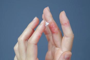
One of the dermatological findings to first gain traction among COVID-19-related manifestations is chilblains-like lesions, or commonly referred to as “COVID toes” or pseudo-chilblains. Chilblains, also known as perniosis, is a localized vasculopathy that involves vasoconstriction of small arterioles and venules triggered by cold, most commonly affecting those with chronic exposure to low temperatures, damp exposure or poor peripheral circulation, in children, young women, and elderly patients. Lesions present as either single or multifocal erythematous, purplish edematous papules, nodules, macules or less commonly bullae, that may be pruritic, tender or accompanied by a paresthesia, commonly on the extremities such as the fingers, toes (Figure 1), and less commonly on nose and ears
Variable COVID-19 testing results among reported patients, however, has sparked some debate on whether pseudo-chilblains are truly a sign of COVID-19 or rather secondary to quarantine-related lifestyle changes. Cordoro and colleagues6 reported clustered cases of pseudo-chilblains appearing within 1 week among 2 groups of siblings aged 12 to 17 years old, in Northern California, in the context of either mild viral URI symptoms (subjective fever, rhinorrhea) or contact with symptomatic persons 1-2 weeks before rash onset. However, SARS-CoV-2 PCR and IgG, IgM antibodies were negative for all patients. Similarly, a combined study of prospective testing in Eastern Spain and retrospective literature review of acral cases in both adults and children found only 13 out of 88 (14.8%) tested cases were positive for COVID-19.5 PCR/serology testing of 38 children (median age 13.5 years) with new onset pseudo-chilblains at a tertiary care university hospital in Italy found all patients negative for RT-PCR and IgM, IgG, IgA antibodies
Quarantine-related habits may contribute to the rise in primary chilblains cases, but the association with COVID-19 cannot be entirely ruled out by the absence of positive PCR or serology. Initial viral testing is biased towards patients with URI symptoms, the timing of retrospective testing is variable, and much is still unknown about COVID-19 antibodies - including the percentage of who acquires these antibodies and the duration of positive serology.
Erythema multiforme-like lesions also presented in a case series of adolescents (aged 11 to 17 years), in conjunction with pseudo-chilblains and possible COVID-19 relation.14 Erythema multiforme (EM) is a hypersensitivity syndrome with distinct mucocutaneous manifestations in the setting of infection. It is characterized by acute onset of target lesions (sharply demarcated round plaques with 3 zones: outermost erythematous ring, middle pale zone, central dusking with possible blistering or crusting), that affects mostly the extremities and are most commonly triggered among children and young adults by viral infections, namely herpes simplex virus (HSV) and by Mycoplasma pneumoniae.
The authors have proposed that vascular damage clinically manifesting as pseudo-chilblains may represent a late antigen-antibody reaction at a time when viral load is already low. This theory is supported by the relatively late onset of lesions and significant number of negative nasopharyngeal PCRs. In addition, these patients also seem to have an overall milder disease presentation.
The Pediatric Dermatology Research Alliance COVID-19 Response Task Force (PDCRTF) has developed an open registry for cases to study and better understand this phenomenon.
Other vascular related skin manifestations such as livedo-reticularis and purpuric or petechial eruptions have been reported in adults and were felt to be a thrombotic phenomenon related to viral infection, but a literature search has not revealed such cases among children up to date of this publication except for one case of petechiae presenting in relation to other morbilliform and annular lesions discussed later.
Nonspecific viral exanthems
A series of cases highlights a varicella-like exanthem associated with COVID-19 among adult patients, particularly in Italy, who developed a monomorphic papulovesicular rash of the trunk that progressed to crusting over a few days with histology consistent with viral infection.
Aside from the more widespread truncal papulovesicular lesions, there are also reports of more localized eruptions. Klimach et al19 described a COVID19 positive case of a boy aged 13 years, in the United Kingdom with an erythematous papular eruption involving the axillae and feet, as well as erythematous macules with associated petechiae and annular lesions on the distal lower extremities. The skin findings developed either before or within 24 hours of onset of mild flu-like symptoms (fever, myalgia and headache), and resolved within 2 weeks.
Of note, an early pruritic morbiliform eruption, with onset concomitant with other COVID-19 symptoms and lasting 3-10 days has been described in adults infected with SARSCoV-222 In children, morbiliform eruptions have been most commonly described in the context of MIS-C, as discussed below. Although urticarial eruptions have been described in adults, to our knowledge at time of this publication, no isolated cases have been reported in children in relation to COVID-19.
Similar to the presentation of pediatric patients with chilblains-like lesions, these viral exanthems seem to affect children who are generally asymptomatic or with mild preceding URI symptoms. Notably, the onset of these generalized cutaneous findings tends to be earlier than onset of vascular-related findings, presenting within a few days of any other viral symptoms.
Multisystem inflammatory syndrome in children (MIS-C) and KD-like
Viral infections among children are generally self-limiting, but in rare cases can trigger multisystem inflammation and severe sequelae. SARS-CoV-2 seems to be no exception, and although current literature demonstrates the majority of infections among children to be asymptomatic or mild-moderate cases, there are temporal relationships to KD-like presentations to COVID-19, including in Italy where one hospital saw a 30-fold increase in the rate of KD-like presentation during the COVID-19 pandemic.
Notably, reports of MIS-C and KD-like have demonstrated large prevalence of mucocutaneous involvement in these patients, with some presenting as one of the primary or most prominent initial symptoms of the disease course. Patients can present with bilateral non-purulent conjunctival injection and oropharyngeal changes including diffuse hyperemia, strawberry tongue, and lip fissures. Cutaneous presentations can also involve hand-foot erythema and edema progressing later to desquamation, and diffuse exanthema with reports of various potential morphologies including either morbiliform, urticarial, EM-like, scarlatiniform, or pustular lesions (Figure 2).
A large retrospective study conducted by Feldstein and colleagues
Among patients with recorded COVID-19 symptoms before MIS-C onset, the median time interval measured in one study was 25 days.26 Additionally, the emergence of reported MIS-C cases during the descent of the COVID-19 epidemic in those locations suggests MIS-C has a delayed onset after SARS-CoV-2 infection.26 Concerning the association between COVID-19 and MIS-C or KD-like presentation, it is important to consider the temporal relationship of generalized exanthems appearing as one of the most common, or even first clinical manifestations of these multi-system inflammatory responses.
Conclusion
A variety of dermatologic findings have been reported thus far amongst pediatric patients in relation to COVID-19, best categorized into chilblains-like lesions, viral exanthem variations (morbiliform, varicella-like), and mucocutaneous involvement as part of KD-like and MIS-C. Pseudo-chilblains and varicella-like eruptions tend to present in asymptomatic or mildly symptomatic patients within 1-2 weeks of respiratory symptoms or contact with infected persons and self-resolve, with some reports of histological evidence of inflammatory infiltration and SARS-CoV-2 positive immunohistochemistry in patients regardless of PCR or serological testing. On the other hand, KD-like and MIS-C patients tend to present later in the course of COVID-19 infection, with predominant mucocutaneous involvement (generalized exanthem, cheilitis) that can even be the early feature on initial presentation. The temporal and causal relationships between COVID-19 and each of these cutaneous manifestations are still unclear due to variable viral testing and follow-up, and more in-depth studies are needed to understand the correlation.
Diagnosing of these cutaneous findings can be challenging as they are nonspecific and can also be seen in the context of other viral or bacterial infections, drug reactions, or have idiopathic etiology. Therefore, when evaluating patients with such cutaneous findings, COVID-19 should be suspected but it is important to still consider and rule alternative causes.29
References
1. The World Health Organization (WHO). Coronavirus Disease (COVID-19) Dashboard. Accessed July 28, 2020. Available at:
2. Guan WJ, Ni ZY, Hu Y, et al. Clinical characteristics of coronavirus disease 2019 in China. N Engl J Med. 2020;382(18):1708-1720. doi:10.1056/NEJMoa2002032
3. Dong Y, Mo X, Hu Y, et al. Epidemiology of COVID-19 among children in China. Pediatrics. 2020;145(6):e20200702. doi:10.1542/peds.2020-0702
4. Stokes EK, Zambrano LD, Anderson KN, et al. Coronavirus disease 2019 case surveillance —United States, January 22–May 30, 2020. MMWR Morb Mortal Wkly Rep 2020;69:759–765. doi: 10.15585/mmwr.mm6924e2
5. Simon TD, Soep JB, Hollister JR. Pernio in pediatrics. Pediatrics. 2005;116(3):e472-e475. doi:10.1542/peds.2004-2681
6. Cordoro KM, Reynolds SD, Wattier R, McCalmont TH. Clustered cases of acral perniosis: clinical features, histopathology, and relationship to COVID-19. Pediatr Dermatol. 2020;37(3):419-423. doi:10.1111/pde.14227
7. Caselli D, Chironna M, Loconsole D, et al. No evidence of SARS-Cov-2 infection by PCR or serology in children with pseudochilblain [published online ahead of print, 2020 Jul 1]. Br J Dermatol. July 1, 2020. Epub ahead of print. doi:10.1111/bjd.19349
8. Neri I, Virdi A, Corsini I, et al. Major cluster of paediatric 'true' primary chilblains during the COVID-19 pandemic: a consequence of lifestyle changes due to lockdown. J Eur Acad Dermatol Venereol. June 13, 2020. Epub ahead of print. doi:10.1111/jdv.16751
9. Caselli D, Chironna M, Loconsole D, et al. No evidence of SARS-Cov-2 infection by PCR or serology in children with pseudochilblain. Br J Dermatol. July 1, 2020. Epub ahead of print. doi:10.1111/bjd.19349
10. Sethuraman N, Jeremiah SS, Ryo A. Interpreting diagnostic tests for SARS-CoV-2 ]. JAMA. May 6,2020. Epub ahead of print. doi:10.1001/jama.2020.8259
11. El Hachem M, Diociaiuti A, Concato C, et al. A clinical, histopathological and laboratory study of 19 consecutive Italian paediatric patients with chilblain-like lesions: lights and shadows on the relationship with COVID-19 infection. J Eur Acad Dermatol Venereol. May 31, 2020. Epub ahead of print. doi:10.1111/jdv.16682
12. Colmenero I, Santonja C, Alonso-Riaño M, et al. SARS-CoV-2 endothelial infection causes COVID-19 chilblains: histopathological, immunohistochemical and ultraestructural study of 7 paediatric cases. Br J Dermatol. June 20, 2020. Epub ahead of print. doi:10.1111/bjd.19327
13. Weston WL, Morelli JG. Childhood pernio and cryoproteins. Pediatr Dermatol. 2000;17(2):97-99. doi:10.1046/j.1525-1470.2000.01722.
14. Torrelo A, Andina D, Santonja C, et al. Erythema multiforme-like lesions in children and COVID-19. Pediatr Dermatol. 2020;37(3):442-446. doi:10.1111/pde.14246
15. Zoghaib S, Kechichian E, Souaid K, Soutou B, Helou J, Tomb R. Triggers, clinical manifestations, and management of pediatric erythema multiforme: A systematic review. J Am Acad Dermatol. 2019;81(3):813-822. doi:10.1016/j.jaad.2019.02.057
16. Landa N, Mendieta-Eckert M, Fonda-Pascual P, Aguirre T. Chilblain-like lesions on feet and hands during the COVID-19 Pandemic. Int J Dermatol. 2020;59(6):739-743. doi:10.1111/ijd.14937
17. Galván Casas C, Català A, Carretero Hernández G, et al. Classification of the cutaneous manifestations of COVID-19: a rapid prospective nationwide consensus study in Spain with 375 cases. Br J Dermatol. 2020;183(1):71-77. doi:10.1111/bjd.19163
18. Pediatric Dermatology Research Alliance. COVID acral ischemia/perniosis in children. Accessed on July 16, 2020.
19. Klimach A, Evans J, Stevens J, Creasey N. Rash as a presenting complaint in a child with COVID-19. Pediatr Dermatol. June 11, 2020. Epub ahead of print. doi:10.1111/pde.14257
20. Marzano AV, Genovese G, Fabbrocini G, et al. Varicella-like exanthem as a specific COVID-19-associated skin manifestation: multicenter case series of 22 patients. J Am Acad Dermatol. 2020;83(1):280-285. doi:10.1016/j.jaad.2020.04.044
21. Genovese G, Colonna C, Marzano AV. Varicella-like exanthem associated with COVID-19 in an 8-year-old girl: A diagnostic clue? Pediatr Dermatol. 2020;37(3):435-436. doi:10.1111/pde.14201
22. Gisondi P, PIaserico S, Bordin C, Alaibac M, Girolomoni G, Naldi L. Cutaneous manifestations of SARS-CoV-2 infection: a clinical update. J Eur Acad Dermatol Venereol. June 25, 2020. Epub ahead of print. doi:10.1111/jdv.16774
23. Kaya G, Kaya A, Saurat JH. Clinical and histopathological features and potential pathological mechanisms of skin lesions in COVID-19: review of the literature. Dermatopathology (Basel). 2020;7(1):3-16. doi:10.3390/dermatopathology7010002
24. Verdoni L, Mazza A, Gervasoni A, et al. An outbreak of severe Kawasaki-like disease at the Italian epicentre of the SARS-CoV-2 epidemic: an observational cohort study. Lancet. 2020;395(10239):1771-1778. doi:10.1016/S0140-6736(20)31103-X
25. World Health Organization (WHO). Multisystem inflammatory syndrome in children and adolescents temporally related to COVID-19 – scientific brief. Published May 15, 2020. Accessed July 16, 2020.
26. Feldstein LR, Rose EB, Horwitz SM, et al. Multisystem inflammatory syndrome in US children and adolescents. N Engl J Med. June 29, 2020. Epub ahead of print. doi:10.1056/NEJMoa2021680
27. Jones VG, Mills M, Suarez D, et al. COVID-19 and Kawasaki disease: novel virus and novel case. Hosp Pediatr. 2020;10(6):537-540. doi:10.1542/hpeds.2020-0123
28. Labé P, Ly A, Sin C, et al. Erythema multiforme and Kawasaki disease associated with COVID-19 infection in children [published online ahead of print, 2020 May 26]. J Eur Acad Dermatol Venereol. 2020;10.1111/jdv.16666. doi:10.1111/jdv.16666
29. Bursal Duramaz B, Yozgat CY, Yozgat Y, Turel O. Appearance of skin rash in pediatric patients with COVID-19: three case presentations. Dermatol Ther. May 15, 2020. Epub ahead of print. doi:10.1111/dth.13594
Articles in this issue
over 5 years ago
Bad to the bone: A case of altered mental statusover 5 years ago
COVID-19 and the future of vaccinationsover 5 years ago
Utilizing the Pediatric Symptom Checklistover 5 years ago
Childhood wheezing may protect against future allergyover 5 years ago
Consider the foreskin for syndactyly repairover 5 years ago
Is botulinum toxin effective for treating children’s migraines?Newsletter
Access practical, evidence-based guidance to support better care for our youngest patients. Join our email list for the latest clinical updates.


