
|Articles|August 31, 2008
Eruptive Xanthoma in a Teenager
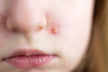
Sixteen-year-old African American girl with pruritic, painful lesions on both thighs that progressively worsened over 2 weeks and later spread to the trunk and upper and lower extremities.
Advertisement
Figure
Figure
>HISTORY
Sixteen-year-old African American girl with pruritic, painful lesions on both thighs that progressively worsened over 2 weeks and later spread to the trunk and upper and lower extremities. Over-the-counter 1% hydrocortisone cream and oral diphenhydramine (25 mg) provided no relief. Patient denied change in clothes, lotions, detergents, or soaps; and recent shaving, swimming, hot tub use, new medications, or sick contacts. No fever, chills, malaise, arthralgia, myalgia, cough, coryza, congestion, or recent illness.
Patient had type 2 diabetes mellitus, hypertension, hyperlipidemia, and hypothyroidism. Medications included metformin, 1000 mg bid; glipizide, 5 mg/d; glargine, 40 units/d; rosiglitazone, 4 mg/d; simvastatin, 20 mg/d; fenofibrate, 48 mg/d, and lisinopril 2.5 mg bid. No family history of hyperlipidemia.
PHYSICAL EXAMINATION
Severely obese patient (body weight, 112 kg [247 lb]; body mass index, 38.5) with diffuse, 1- to 2-mm, discrete, tender papules with a yellow hue on the trunk and bilateral upper and lower extremities, mainly on the extensor surfaces. No erythema, drainage, or induration.
WHAT'S YOUR DIAGNOSIS?
ANSWER: ERUPTIVE XANTHOMA
Eruptive xanthoma is a cutaneous eruption in patients with grossly elevated triglyceride levels. This patient's laboratory results included the following levels:
- Triglycerides, 8250 mg/dL (normal, 0 to 149 mg/dL).
- Total cholesterol, 751 mg/dL (normal, 0 to 199 mg/dL).
- High-density lipoprotein, 79 mg/dL (normal, 40 to 59 mg/dL).
- Hemoglobin A1C, 11.4% (normal, less than 6%).
- Random glucose, 250 mg/dL (normal, 60 to 140 mg/dL).
- Thyroid-stimulating hormone, 12 mIU/L, (normal, 0.35 to 5.50 mIU/L).
A punch biopsy of the lesions showed foamy histiocytic infiltrates in the dermis, consistent with eruptive xanthoma.
Xanthoma pathophysiology is similar to that of atherosclerosis formation in blood vessels.1 No specific triglyceride level is associated with the development of eruptive xanthoma.2-6 This patient's eruptive xanthoma was likely caused by uncontrolled type 2 diabetes mellitus, obesity, and hypothyroidism, in addition to the hypertriglyceridemia. Other secondary causes of eruptive xanthoma include familial hyperlipidemia (Fredrickson types I, IV, and V), metabolic diseases (lysosomal storage diseases and type I glycogen storage disease, or von Gierke disease), alcohol ingestion, chronic renal failure, nephrotic syndrome, pancreatitis, and biliary cirrhosis.7 Eruptive xanthoma may result from medications that elevate lipid levels, such as estrogens, corticosteroids, miconazole, isotretinoin, and etretinate. 7 The lesions may also develop despite normal lipid levels in pregnant patients and in patients with altered lipoprotein content or structure, paraproteinaemia, hematopoietic diseases (such as histiocytosis and myeloma), edema, acquired lipodystrophy, and local trauma.7
Despite the lack of a family history of hyperlipidemia, familial hyperlipidemia must be considered in patients with such high lipid levels. Diagnosis of familial hypercholesterolemia can be made in patients with a serum total cholesterol level of greater than 259 mg/dL and/or a lowdensity lipoprotein (LDL) level of 193 to 232 mg/dL who have xanthomas, or in patients with an LDL level of greater than 232 mg/dL alone.8 However, secondary causes must be ruled out. (This patient did not meet these criteria because of her history of type 2 diabetes mellitus, obesity, and hypothyroidism.) Familial-associated xanthoma formation is most often consistent with Fredrickson types I and V familial hyperlipidemia.9
The following conditions may be considered in the differential diagnosis of eruptive xanthoma:
- Varicella. This was unlikely in this patient because of the absence of malaise and a fever prodrome. In addition, her lesions were not clear vesicles on an erythematous base ("dew drop on a rose petal"), characteristic of varicella. The rash also did not appear to be in varying stages of formation or healing.
- Folliculitis. This is usually a self-limited condition and is commonly associated with shaving, exposure to hot temperatures and humidity, or breakage in the skin surface (via scratching) where hair follicles are present. Typically, the lesions are more papular and pustular with surrounding erythema and induration.10
- Sweet syndrome. This typically occurs in women aged 30 to 50 years, usually after an upper respiratory tract infection. The illness is characterized by fever (temperature of greater than 38°C [100.4°F]) with abrupt onset of erythematous plaques or nodules. The histopathology of the lesions is consistent with dense neutrophilic infiltrate without evidence of leukocytoclastic vasculitis.11
Although the duration of eruptive xanthoma varies widely, the rash typically resolves as the triglyceride levels decrease.2-6 Treatment includes rapid control of the blood sugar and triglyceride levels. Blood sugar can be controlled acutely with the initiation of insulin or the adjustment to an existing insulin regimen with close follow-up. Hypertriglyceridemia can be managed with initiation or titration of existing fenofibrates. The underlying medical conditions of hypothyroidism, diabetes, and hyperlipidemia also need to be addressed.
This patient has been nonadherent to her medication regimen, dietary changes, and follow-up appointments despite multiple phone and mail reminders. At her last visit, 6 months after presentation, the xanthomas were still present.
References:
- Kinoshita M, Kawamura M, Fujita M, et al. Enhanced susceptibility of LDL to oxidative modification in a CTX patient: role of chenodeoxycholic acid in xanthoma formation. J Atheroscler Thromb. 2004;11:167-172.
- Shinozaki S, Itabashi N, Rokkaku K, et al. Diabetic lipemia with eruptive xanthomatosis in a lean young female with apolipoprotein E4/4. Diabetes Res Clin Pract. 2005;70:183-192.
- Nayak KR, Daly RG. Images in clinical medicine. Eruptive xanthomas associated with hypertriglyceridemia and new-onset diabetes mellitus. N Engl J Med. 2004;350:1235.
- Naik NS. Eruptive xanthomas. Dermatol Online J. 2001;7:11.
- Park JR, Jung TS, Jung JH, et al. A case of hypothyroidism and type 2 diabetes associated with type V hyperlipoproteinemia and eruptive xanthomas. J Korean Med Sci. 2005;20:502-505.
- Roller E, Schulte KW, Hengge U, et al. Eruptive xanthomas [in German]. Hautarzt. 2004;55:978-980.
- Tang WK. Eruptive xanthoma. Case reports. Hong Kong Dermatol Venereol Bull. 2001;9:172-175.
- Koivisto PV, Koivisto UM, Miettinen TA, Kontula K. Diagnosis of heterozygous familial hypercholesterolemia. DNA analysis complements clinical examination and analysis of serum lipid levels. Arterioscler Thromb. 1992;12: 584-592.
- Fauci AS, Braunwald E, Kasper DL, et al, eds. Harrison's Principles of Internal Medicine. 17th ed. New York: McGraw Hill; 2008:2419.
- Stulberg DL, Penrod MA, Blatny RA. Common bacterial skin infections. Am Fam Physician. 2002;66:119-124.
- Cohen PR. Sweet's syndrome-a comprehensive review of an acute febrile neutrophilic dermatosis. Orphanet J Rare Dis. 2007;2:34.
Advertisement
Related Content
Advertisement


Advertisement
Advertisement
Trending on Contemporary Pediatrics
1
FAQ: Which children with early puberty actually need testing and treatment?
2
ACIP dropped MMRV coverage. New data show it was a lifeline for VFC-eligible kids
3
FDA grants tegacorat orphan, rare pediatric disease status for DMD
4
FDA clears IND for investigational SUMF1 gene therapy in multiple sulfatase deficiency
5