
|Articles|September 1, 2006
- Consultant for Pediatricians Vol 5 No 9
- Volume 5
- Issue 9
Ichthyosis Vulgaris in a 7-Month-Old Boy
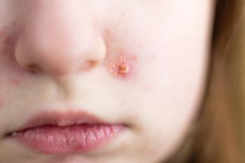
Seven-month-old boy with hyperpigmented lesions on his legs. Lesions first noted at 4 months of age. His 32-year-old father and 58-year-old grandfather had similar lesions on their legs.
Advertisement
HISTORY
Seven-month-old boy with hyperpigmented lesions on his legs (
PHYSICAL EXAMINATION
Brown, polygonal scales present on both shins. No other notable findings. Palms and soles normal. Both testicles in scrotum. No corneal opacity or atopic dermatitis.
"WHAT'S YOUR DIAGNOSIS?"
ANSWER: ICHTHYOSIS VULGARIS
The term ichthyosis is derived from the Greek word ichthys, which means fish, and was chosen because the lesions have the appearance of fish scales. Ichthyosis is a disorder of cornification characterized by the development of dry, rectangular or polygonal scales. Ichthyosis is caused by altered profilaggrin expression, which leads to scaling and desquamation.Over 95% of those with the problem have ichthyosis vulgaris-the mildest form.1 Synonyms for ichthyosis vulgaris include ichthyosis simplex and autosomal dominant ichthyosis.
EPIDEMIOLOGY
The prevalence of ichthyosis vulgaris varies by race; in the white population the prevalence is estimated to be about 0.3% to 0.4%,2 and in the East Asian population, the prevalence is approximately 2.3%.3 Both sexes are affected equally.
GENETICS
Ichthyosis vulgaris is transmitted as an autosomal dominant trait with variable phenotypic expression between and within families.1 The gene locus of ichthyosis vulgaris might reside on chromosome 1q22.2 The profilaggrin gene in this region is part of a cluster of genes that encodes for structural proteins expressed in the terminally differentiating epidermis.
PATHOGENESIS
Profilaggrin is synthesized in the granular layer of the epidermis. Profilaggrin is stored in keratohyaline granules in a highly phosphorylated form4 and undergoes various post-translational modifications to become filaggrin, a filament-aggregating protein.1 Filaggrin is proteolyzed and metabolized into free amino acids that might play a critical role as water-binding compounds in the upper stratum corneum.Profilaggrin, filaggrin, and keratohyaline granules are decreased or absent in the epidermis of persons with ichthyosis.4,5 The primary genetic defect might be a factor that reduces profilaggrin and filaggrin synthesis.6
CLINICAL MANIFESTATIONS
Management Hydration of the skin and prevention of evaporation are important. Bathing once or twice daily in warm water for approximately 5 to 10 minutes helps to hydrate the skin. Afterward, the body should be gently patted dry to minimize trauma to the affected areas. A moisturizing cream or ointment should be immediately applied to minimize evaporation and to keep the skin soft and flexible. Frequent application of a moisturizing agent throughout the day helps to maintain a high level of hydration of the stratum corneum. Preparations that contain urea and alpha-hydroxy acids, such as lactic acid or pyruvic acid, are particularly effective hydration agents.
8
The lesions are not usually present at birth but appear in most patients during the first year of life and in the vast majority by age 5 years. The scaling is symmetric and usually intensifies until puberty and subsequently decreases with age. The color of the fine, fish-like scales varies from white to dirty gray to brown.1 In general, darker scales are seen in dark-skinned persons.1 The lesions can vary from barely visible roughness and dryness to strong, horny plates. The lesions tend to improve during the summer and with increasing humidity and to worsen during the winter,7 when some patients report "lizard-like" skin.
Scaling is most prominent on the extensor aspects of the extremities, particularly the shins. The back is involved more often than the abdomen. The diaper and flexural areas, such as theaxillae and antecubital and popliteal fossae are spared, perhaps because of the higher temperature and humidity in these areas. The scales often curl up at the edges, which imparts a rough feel to the skin. Hyperlinearity of the palms and soles, keratosis pilaris, atopy, and heat intolerance are more common in persons with ichthyosis vulgaris.8,9 Secondary infections can develop in fissures of the hands and feet.
HISTOPATHOLOGY
An attenuated or reduced granular layer, a decreased rete-papillae pattern, and a reduced number of sebaceous glands are characteristic histologic findings.1,7 Ultrastructural studies reveal reduced or absent keratohyaline granules.1,7 These granules have a crumbly or spongy appearance, which reflect defective keratohyaline synthesis.1
DIFFERENTIAL DIAGNOSIS
Ichthyosis vulgaris should be distinguished from X-linked recessive ichthyosis, lamellar ichthyosis, nonbullous congenital ichthyosiform erythroderma, and bullous congenital ichthyosiform erythroderma (epidermolytic hyperkeratosis).
In X-linked recessive ichthyosis, the scales tend to be larger and darker. Sites of predilection include the extremities, preauricular area, neck, and trunk.10 The palms, soles, and face are characteristically spared. Affected persons might have undescended testes and corneal opacities. Female carriers may pre-sent with a fine, silver-light scaling on the legs.11 In contrast to ichthyosis vulgaris, the lesions in X-linked recessive ichthyosis do not significantly diminish with age.
Lamellar ichthyosis-an autosomal recessive disorder-usually presents at birth with aparchment-like collodion membrane (hence the term collodion baby), which desquamates over the next 10 to 14 days.12 Over time, large, dark brown, plate-like scales develop; these are centrally adherent with raised edges, and resemble a suit of armor. Tautness of the facial skin might result in eversion of the eyelids (ectropion) and lips (eclabium). Unlike ichthyosis vulgaris, flexural areas are involved in lamellar ichthyosis and the palms and soles are almost always affected.
Nonbullous congenital ichthyosiform erythroderma, an autosomal recessive disorder, also presents at birth with a collodion membrane. After shedding of the membrane, pronounced erythroderma and fine white scales distinguish this condition from other forms of ichthyosis.
Bullous congenital ichthyosiform erythroderma, an autosomal dominant disorder, usually presents at birth with erosions, large areas of denuded skin, and erythroderma. Over time, blistering and erythroderma diminish and hyperkeratosis develops. Thick brown scales cover most of the skin surface, especially in the flexural areas.
References:
REFERENCES:
1. Okulicz JF, Schwartz RA. Hereditary and acquired ichthyosis vulgaris.
Int J Dermatol.
2003;42:95-98.
2. Zhong W, Cui B, Zhang Y, et al. Linkage analysis suggests a locus of ichthyosis vulgaris on 1q22.
J Hum Genet.
2003;48:390-392.
3. Lei G, Zhang Y, Hu Y. Investigation on the prevalence of ichthyosis in Sichuan Province
. Chin J Dermatol.
1992;25:105-106.
4. Fleckman P, Brumbaugh S. Absence of the granular layer and keratohyalin define a morphologically distinct subset of individuals with ichthyosis vulgaris.
Exp Dermatol.
2002;11:327-336.
5. Sybert VP, Dale BA, Holbrook KA. Ichthyosis vulgaris: identification of a defect in synthesis of filaggrin correlated with an absence of keratohyaline granules.
J Invest Dermatol.
1985;84:191-194.
6. Nirunsuksiri W, Zhang SH, Fleckman P. Reduced stability and bi-alleic, coequal expression of profilaggrin mRNA in keratocytes cultured from subjects with ichthyosis vulgaris.
J Invest Dermatol.
1998;100:854-861.
7. Richard G, Moss C, Traupe H, et al. Ichthyosis and disorders of cornification. In: Schachner LA, Hansen RC, Happle R, et al, eds.
Pediatric Dermatology.
Philadelphia: Mosby; 2003:385-445.
8. Leung AK, Kao CP. Keratosis pilaris.
Consultant For Pediatricians.
2004;3: 188-191.
9. Richard G, Ringpfeil F. Ichthyosis, erythrokeratomas and related disorders. In: Bolognia J, Jorizzo JL, Rapini RP, eds.
Dermatology.
Philadelphia: Mosby; 2003:775-808.
10. Okano M, Kitano Y, Yoshikawa K, et al. X-linked ichthyosis and ichthyosis vulgaris: comparison of their clinical features based on biochemical analysis.
Br J Dermatol.
1988;119:777-783.
11. Voss M. Clinical picture of X chromosome recessive ichthyosis.
Dermatol Monatsschr.
1985;171:25-37.
12. Leung AK. Collodion baby.
Consultant For Pediatricians.
2004;3:233.
Articles in this issue
almost 20 years ago
Botulinum Toxin Therapy in Children:almost 20 years ago
Case In Point: An Unusual Case of Ileal-Ileo Intussusceptionalmost 20 years ago
Genetic Disorders: Tetany in a 9-Year-Old Girlalmost 20 years ago
A Collage of Infectious Diseases in Childrenalmost 20 years ago
Erratum: Update on treatment of primary syphilisalmost 20 years ago
Pediatric Migraine: Clinical Pearls in Diagnosis and Therapyalmost 20 years ago
Musculoskeletal Clinics: 16-Year-Old Camper With Tibial Painalmost 20 years ago
Photoclinic: Corneal Abrasionalmost 20 years ago
Case In Point: Spontaneous Pneumothorax in a Teenage Boyalmost 20 years ago
Pediatric Musculoskeletal Infections: Combating the Major PathogensAdvertisement
Related Content
Advertisement


Advertisement
Advertisement
Trending on Contemporary Pediatrics
1
ACIP dropped MMRV coverage. New data show it was a lifeline for VFC-eligible kids
2
FAQ: Which children with early puberty actually need testing and treatment?
3
Douglas Mack, MD, says THRIVE study could change early peanut allergy management
4
FDA clears IND for investigational SUMF1 gene therapy in multiple sulfatase deficiency
5