
|Articles|March 1, 2008
Pityriasis Rosea in a 17-Year-Old Girl
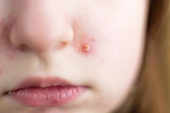
17-Year-old female with a salmon-pink oval patch on her back. Three days after the patch appeared, a generalized eruption developed on her back. Eruption slightly pruritic. No antecedent upper respiratory tract infection. No prodromal symptoms.
Advertisement
Figure
Figure
Figure>
HISTORY
17-Year-old female with a salmon-pink oval patch on her back. Three days after the patch appeared, a generalized eruption developed on her back. Eruption slightly pruritic. No antecedent upper respiratory tract infection. No prodromal symptoms.
Past health unremarkable. Patient not taking any medication. No history of sexually transmitted diseases and no behavioral risk factors for them.
PHYSICAL EXAMINATION
Patient looks healthy overall: free of features of acute or chronic illness. Apart from papulosquamous skin eruptions, physical findings are normal. In particular, no abnormality of oral or ocular mucous membrane, nails, external auditory canal, or vulvar or perianal skin.
ANSWER: PITYRIASIS ROSEA
Pityriasis rosea is an acute, self-limited papulosquamous dermatosis-a skin condition with bumps and lots of scaling-that primarily affects children and young adults. The condition was first described by Robert Willan in 1798. The term "pityriasis (scaly) rosea (pink)" was coined by Camille Melchior Gibert in 1860.1 Synonyms include roseola annulata, pityriasis maculata et circinata, and pityriasis rubra aigu dissmin.
CLINICAL MANIFESTATIONS
The most common presenting sign-found in 59% to 90% of patients2,3-is a herald or "mother" patch, which begins as a smooth, erythematous macule or papule; it expands over 1 to 2 weeks and then forms a round or oval, erythematous scaly lesion that can range anywhere from 1 to 10 cm in diameter.4 Herald patches occur in other conditions as well, notably in the commonly drug-induced eruption of erythema multiforme. Thus the mere presence of such a patch is not diagnostic of pityriasis rosea.
The herald patch of pityriasis rosea is usually found on the trunk and less often on the neck and proximal extremities.5 Multiple herald patches are found in approximately 5% of patients.6 A mild prodrome consisting of headache, fever, malaise, anorexia, sore throat, and arthralgia is present in about 5% of patients and might reflect the effects of a viral illness that has triggered the skin exanthem. However, virus has never been recovered from the lesions of pityriasis rosea.5
A generalized, bilateral, symmetrical eruption develops in up to 21 days after the appearance of the herald patch and continues to erupt in crops over the next 10 to 21 days.7 For whatever reason, 21 days seems to be a magic interval in pediatric dermatology. (It is also the longest usual incubation period of chickenpox.) Typical lesions are 5 to 10 mm, pinkish to brown, with a delicate collarette of scale at the periphery. The long axes are along the skin lines of cleavage (Langer lines).2,7
The distribution on the back thus forms a "Christmas tree" or "fir tree" appearance that can be extremely striking. Involvement of the face and scalp is unusual except in black persons.7
Rarely, the palms and soles may be affected.4,7 In some children-particularly African-Americans-there is an inverse distribution, with lesions mostly on the face and in the axillary and inguinal areas. Oral lesions are uncommon and include punctate hemorrhages, vesicles, and bullae. Pruritus is present in most cases.6,7 Significant systemic manifestations are characteristically absent. Resolution usually requires 2 to 12 weeks.3
In dark-skinned persons, the lesions tend to be more papular and hyperpigmented.5,8 Also-and similar to the effects of other inflammatory skin processes- post-inflammatory hyperpigmentation or hypopigmentation persists after the resolution of the acute lesions more often in darker- skinned persons.7
EPIDEMIOLOGY
Approximately 75% of cases occur in persons between the ages of 10 and 35 years.6 The peak incidence is during adolescence. The condition is rare in children younger than 2 years. There is a slight female predominance.6,9 Most cases occur in the spring and autumn. The disease occurs worldwide. The average annual incidence rate is estimated at 170 per 100,000 person- years in the United States.9
PATHOGENESIS AND ETIOLOGY
A viral etiology has been proposed because of the seasonal variation, clustering in communities, and the symptoms of antecedent upper respiration tract infection (presumed to be viral) found in up to 20% of affected patients.3 Human herpesvirus 7 and, to a lesser degree, human herpesvirus 6 have been implicated, but the causative role is still controversial.10,11 Histologically, focal parakeratosis, spongiosis, and a superficial lymphohistiocytic perivascular infiltrate might be present.10
DIFFERENTIAL DIAGNOSIS
The diagnosis is clinical and based on characteristic findings, and there is no laboratory test to confirm or exclude the diagnosis; nor is histopathological examination of a skin biopsy specimen necessarily conclusive. The differential diagnosis includes tinea corporis, guttate psoriasis, secondary syphilis, nummular eczema, pityriasis lichenoides, pityriasis versicolor, and drug eruption. Pityriasis rosea-like drug eruptions can be caused by penicillamine, captopril, bismuth, ketotifen, barbiturates, and isotretinoin.12,13 A toxic rather than allergic-type pityriasis rosea-like eruption has also been reported following topical application of mustard oil.14
TREATMENT
The patient should be reassured that the rash will resolve spontaneously within 12 weeks, and that it rarely persists for more than 5 months.8 Pruritus can be managed with a topical corticosteroid or an oral antihistamine. Treatment options for severe cases include erythromycin and UVB phototherapy.4,6 Azithromycin is not effective in the treatment of this condition.15
References:
- Gibert CM. Traite Pratique Des Maladies de la Peau et de la Syphilis. 3rd ed. Paris: H. Plon; 1860:402.
- Leung AK, Wong BE, Chan PY, et al. Pityriasis rosea. Resident Staff Physician. 1997;43:109.
- Bernardin RM, Ritter SE, Murchland MR. Papular pityriasis rosea. Cutis. 2002;70:51-55.
- Leung AK, Robson WL, Woo TY. Pityriasis rosea. In: Lang F, ed. The Encyclopedia of Molecular Mechanisms of Disease. Berlin: Springer-Verlag. In press.
- Wood GS, Reizner G. Pityriasis rosea. In: Bolognia JL, Jorizzo JL, Rapini RP, eds. Dermatology. Philadelphia: Mosby; 2003:158-160.
- González LM, Allen R, Janniger CK, Schwartz RA. Pityriasis rosea: an important papulosquamous disorder. Int J Dermatol. 2005;44:757-764.
- Amer A, Fischer H, Li X. The natural history of pityriasis rosea in black American children: how correct is the "classic" description? Arch Pediatr Adolesc Med. 2007;161:503-506.
- Amer A, Fischer H. Pityriasis rosea. Consultant Pediatricians. 2006;5:176-178.
- Bianca S, Ingegnosi C, Ciancio B, et al. Pityriasis rosea in pregnancy. Reprod Toxicol. 2007;24:277-278.
- Drago F, Vecchio F, Rebora A. Use of high-dose acyclovir in pityriasis rosea. J Am Acad Dermatol. 2006;54:82-85.
- Dyer JA. Childhood viral exanthems. Pediatr Ann. 2007;36:21-29.
- Atzori L, Pinna AL, Ferreli C, Aste N. Pityriasis rosea-like adverse reaction: review of the literature and experience of an Italian drug-surveillance center. Dermatol Online J. 2006;12:1.
- Hanjani NM, Rencic A, Whitmore SE. Pityriasis rosea-like eruption due to bismuth. Cutis. 2006;77:166-168.
- Zawar V. Pityriasis rosea-like eruptions due to mustard oil application. Indian J Dermatol Venereol Leprol. 2005;71:282-284.
- Amer A, Fischer H. Azithromycin does not cure pityriasis rosea. Pediatrics. 2006;117:1702-1705.
Advertisement
Related Content
Advertisement


Advertisement
Advertisement
Trending on Contemporary Pediatrics
1
FAQ: Which children with early puberty actually need testing and treatment?
2
ACIP dropped MMRV coverage. New data show it was a lifeline for VFC-eligible kids
3
FDA grants tegacorat orphan, rare pediatric disease status for DMD
4
FDA clears IND for investigational SUMF1 gene therapy in multiple sulfatase deficiency
5