- Vol 37 No 8
- Volume 37
- Issue 8
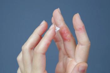
Bully bullae in a toddler spreads across the body
You are asked to evaluate a healthy 19-month-old boy with a history of recurrent blisters on his arms and legs for over a year that have now spread to his face. The blisters scab over quickly, ulcerate, and heal with post-inflammatory pigmentary changes. What's the diagnosis?
The case
You are asked to evaluate a healthy 19-month-old boy with a history of recurrent blisters on his arms and legs for over a year that have now spread to his face. The blisters scab over quickly, ulcerate, and heal with post-inflammatory pigmentary changes.
Diagnosis: Chronic bullous disease of childhood (CBDC)
Discussion
Chronic bullous disease of childhood, also known as linear IgA bullous dermatosis (LABD), is an uncommon disease that occurs in children between 6 months and 10 years.1 Chronic bullous disease of childhood presents with expanding annular erythematous plaques with blisters at the periphery, resulting in the string of pearls sign. Early lesions can develop in the flexural areas, although this may vary.1 Commonly involved areas include the face, abdomen, genital area, and extremities.
Chronic bullous disease of childhood is characterized by the linear deposition of IgA along the basement membrane zone (BMZ).1 This leads to neutrophilic infiltrate and subepidermal bullae, often with increased prevalence of eosinophils.1 LABD cannot be definitively diagnosed by light microscopy and should be confirmed with immunofluorescent studies which show linear staining for IgA at the basement membrane zone.1
Conditions associated with LABD include autoimmune diseases, malignancy, and infections,1 with insufficient data to determine the statistical significance of these relationships.2 One known association is drug-induced LABD, especially with vancomycin.2 This condition differs from idiopathic LABD in both distribution and disease course.2
Differential diagnosis
The differential diagnosis for CBDC/LABD is extensive; the two most common conditions that mimic LABD are dermatitis herpetiformis and bullous pemphigoid, which may present with bullae or vesicles in similar distributions.1 Direct immunofluorescence is the gold standard for differentiating between these conditions in the absence of other clinical distinguishers.1 The linear deposition of IgA along the BMZ is pathognomonic for CBDC and LABD; dermatitis herpetiformis is characterized by granular IgA deposition in the dermal papillary tips or along the basement membrane and bullous pemphigoid is characterized by linear IgG deposition along the epidermal BMZ.1 Besides direct immunofluorescence, circulating anti-BMZ IgA antibodies may be found in 60% to 70% of LABD patients, not found in dermatitis herpetiformis or bullous pemphigoid.1
Management
Although CBDC is generally self-limited, it may lead to morbidity and requires systemic treatment.3 The standard of care for treatment is dapsone for controlling blistering without adverse reactions in most cases.3 Dapsone is initiated at low doses (<0.5mg/kg) and increased until symptoms are controlled.3 Adverse effects include hemolysis, especially in patients with glucose-6-phosphate dehydrogenase deficiency.3 Second-line therapies include erythromycin, dicloxacillin, oxacillin, colchicine, and systemic corticosteroids.3
Patient outcome
Chronic bullous disease of childhood was diagnosed clinically. Following bloodwork, this patient started on dapsone 12.5 mg, then 18.75 mg, with improvement of the lesions. A skin biopsy and direct immunofluorescence confirmed the diagnosis
References
- Hull CM, Zone, JJ. Dermatitis herpetiformis and linear IgA bullous dermatosis. Dermatology. 2017;(4):527–537.
- Kuechle, MK, Stegemeir, E, Maynard, B, et al. Drug-induced linear IgA bullous dermatosis: report of six cases and review of the literature. J Am Acad Dermatol. 1994;30(2):187–192. doi: 10.1016/s0190-9622(94)70015-x
- Mintz, EM, Morel, KD. Treatment of chronic bullous disease of childhood. Dermatol Clin. 2011 29(4):699–700. doi: 10.1016/j.det.2011.08.013
Articles in this issue
over 5 years ago
Safe return to school: Part 2over 5 years ago
A fever, liver abnormalities, and pancytopeniaover 5 years ago
Back to school, or back to remote learning?over 5 years ago
COVID-19: Keep smiling!over 5 years ago
Recommendations for compounded hand sanitizers during COVID-19over 5 years ago
New approach to EOS reduces testing and antibioticsover 5 years ago
Safe return to school: A call to actionover 5 years ago
Smartphone apps for weight control?: How to chooseover 5 years ago
Melatonin helps children with ASD overcome insomniaNewsletter
Access practical, evidence-based guidance to support better care for our youngest patients. Join our email list for the latest clinical updates.


