- Consultant for Pediatricians Vol 6 No 12
- Volume 6
- Issue 12
Child With "Bruising"
A 23-month-old Hispanic boy was brought to an emergency department (ED) with ear pain and fever. The family had no other expressed concerns. Physical examination revealed inflammation of 1 tympanic membrane. The child had a temperature of 38.4°C (101.2°F).
A 23-month-old Hispanic boy was brought to an emergency department (ED) with ear pain and fever. The family had no other expressed concerns. Physical examination revealed inflammation of 1 tympanic membrane. The child had a temperature of 38.4°C (101.2°F).
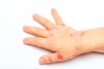
On closer examination, the child had skin discoloration on the chest that looked like bruising. The findings from the remainder of the examination were normal, and there were no other indications of possible abuse. The family could offer no explanation for the bruising. The mother was certain that the discoloration had not been present when she took the child outside to play earlier in the day.
Results of a complete blood cell count, prothrombin time, and partial thromboplastin time were all within normal limits. Child Protective Services was notified of possible nonaccidental injury, and the child was referred to an advocacy center for consultation. The child was seen in consultation within 24 hours of the ED visit.
Do you suspect abuse or a medical cause?
(Answer and discussion on next page.)
Answer: Phytophotodermatitis
On close questioning, the child's mother volunteered that she had been preparing a meal with fresh limes on the day of the ED visit. She recalled that while cooking, she had repeatedly picked up her child without washing her hands and was certain that the child's bare skin had been touched in the process. This detail coupled with the results of the child's physical examination led to a diagnosis of phytophotodermatitis (PPD). The family was cleared of allegations of abuse.
The term "phytophotodermatitis" was first used to describe this condition in 1942 by Dr Robert Klaber.1 Much of what is still considered to be fact about this condition was written in his article. Dermatitis from plant material had been described for many years, but the connection with light--specifically UV light--had not been made. Klaber reviewed a number of case reports and clinical studies to describe the spectrum of the disorder.
Psoralen (from the furocoumarin family) in combination with UV light in the 320- to 380-nm range is the chemical photosensitizer responsible for PPD.2 Cell death and inhibition of mitosis are caused by the cross-linking of DNA with psoralen. Psoralen is present in a number of ordinary foods and in a variety of weeds and grasses that are common in urban areas (Table).3 Cooks, grocers, and gardeners are prone to PPD. There are case reports of PPD caused by airborne plant particulate matter from lawn tools.4
PPD occurs more frequently in the late summer when doses of UV light are high. The heat and humidity of summer months also accentuate the reaction. Typically, the onset of symptoms is within 24 to 48 hours after exposure. The first signs may be painful erythema and local edema. Other reactions may start with hyperpigmentation. More intense reactions may progress to blisters and ultimately to bullae. The degree of reaction is thought to be dose-related and dependent on a patient's skin pigmentation. Persons with light skin are more vulnerable to PPD.
PPD may last weeks to months in certain patients. In some, permanent scarring results. Treatment should be tailored to the intensity of the dermatitis. Patients with mild to moderate symptoms may benefit from high-potency topical corticosteroids. Those with severe manifestations are treated with systemic corticosteroids. For hyperpigmentation, no treatment is necessary, even if it persists for months.
THE KEY POINTS
While this patient's case was straightforward once all the facts were exposed, it illuminates the following key points:
•PPD can be initiated by a vast number of activities and exposures. Children have possible multiple exposures to psoralen on any given day.
•Abuse can often be included in the differential diagnosis in children who present to the ED. Thus, clinicians need to be aware of the conditions that mimic abuse.5,6 PPD can be included in any differential diagnosis in patients who present with burns or bruising.
This patient's case also points out the need for emergent/emergency referral resources to limit the disruption of daily family life by unfounded referrals.7,8 *
References:
REFERENCES:
1.
Klaber R. Phytophotodermatitis.
Br J Dermatol.
1942;54:193-211.
2.
Hipkin CR. Phytophotodermatitis, a botanical view.
Lancet.
1991;338:892-893.
3.
Photosensitivity and photoreactions. In: Hurwitz S, ed.
Clinical Pediatric Dermatology. A Textbook of Skin Disorders of Childhood and Adolescence.
2nd ed. Philadelphia: Saunders; 1993:89-90.
4.
Oakley AM, Ive FA, Harrison MA. String trimmer's dermatitis.
J Soc Occup Med.
1986;36:143-144.
5.
Hill PF, Pickford M, Parkhouse N. Phytophotodermatitis mimicking child abuse.
J R Soc Med.
1997;90:560-561.
6.
Klaber RE. Phytophotodermatitis.
Arch Dis Child.
2006;91:385.
7.
Wallace GH, Makoroff KL, Malott HA, Shapiro RA. Hospital-based multidisciplinary teams can prevent unnecessary child abuse reports and out-of-home placements.
Child Abuse Negl.
2007;31:623-629.
8.
Botash AS, Sveen AR, Frasier LD. Possible burns in 2 children.
Consultant For Pediatricians.
2003;2:334-337.
Articles in this issue
about 18 years ago
Folk Remedy as a Cause of Septicemia in a Child With Leukemiaabout 18 years ago
Neonatal Teethabout 18 years ago
In Bacterial Meningitis, Can Dexamethasone Help?about 18 years ago
PHACE Syndromeabout 18 years ago
Are Late-Preterm Infants Mature Enough?about 18 years ago
Chokingabout 18 years ago
Strawberry Hemangiomaabout 18 years ago
Toddler Who "Caught Psoriasis" at Her Day-Care CenterNewsletter
Access practical, evidence-based guidance to support better care for our youngest patients. Join our email list for the latest clinical updates.


