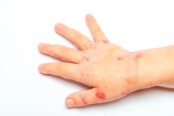
- Consultant for Pediatricians Vol 9 No 6
- Volume 9
- Issue 6
Drug Eruptions: The Benign-and the Life-Threatening
“Drug rash” is a common pediatric complaint in both inpatient and outpatient settings. This term, however, denotes a clinical category and is not a precise diagnosis. Proper identification and classification of drug eruptions in children are important for determining the possibility of-and preventing progression to-internal involvement. Accurate identification is also important so that patients and their parents can be counseled to avoid future problematic drug exposures.
"Drug rash" is a common pediatric complaint in both inpatient and outpatient settings. This term, however, denotes a clinical category and is not a precise diagnosis. Proper identification and classification of drug eruptions in children are important for determining the possibility of-and preventing progression to-internal involvement. Accurate identification is also important so that patients and their parents can be counseled to avoid future problematic drug exposures.
In this article, we provide pertinent information about several cutaneous drug reactions encountered in clinical practice. We emphasize those clinical features that are especially important for clinicians who may be diagnosing and managing these classic eruptions in the pediatric population.
GENERAL APPROACH TO A SUSPECTED DRUG ERUPTION
A detailed history is essential to establish the temporal relationship of the eruption to prior drug exposure and exposure to cross-reacting medications. It is also important to be aware of patterns of drug metabolism, interactions, and toxicities.
NON–LIFE-THREATENING DRUG ERUPTIONS
Figure 1 – The exanthematous drug eruption in this child demonstrates confluent areas of morbilliform erythema with small white areas of sparing.
Exanthematous drug eruption. The most common drug eruptions in children are morbilliform or exanthematous reactions.1 Onset can be anywhere from 1 to 2 weeks following medication administration and may occur or persist even after the responsible drug is discontinued, especially when that drug has a prolonged half-life. The time from medication administration to onset of the drug eruption is reduced with rechallenge.
Note that the likelihood of a reaction occurring is increased in patients who have a coexisting viral infection. Some have hypothesized that the presence of the viral infection may induce the body's immune system to respond to the drug differently than it normally would, thereby increasing the risk of a reaction.2
Culprit drugs. The agents that most frequently cause exanthematous drug eruptions are anticonvulsants and antibiotics. Medications that have been implicated include penicillins,3-7 cephalosporins,3-5 sulfonamides,3-5,7 erythromycin,3 NSAIDs,3,7 barbiturates,3 phenytoin,3,7 carbamazepine,3,7 and benzodiazepines.3
Clinical features. These eruptions consist of erythematous flat macules and papules that begin on the trunk and extend in a symmetrical fashion to become generalized (
Differential diagnosis. In addition to an exanthematous drug eruption, consider viral exanthems with palmoplantar involvement as well as toxic shock syndrome and Kawasaki disease3; however, these last 2 entities are typically accompanied by fever and irritability or other systemic signs, such as hemodynamic instability. It is especially important to distinguish between an exanthematous drug eruption and drug hypersensitivity syndrome, also known as drug reaction with eosinophilia and systemic symptoms (DRESS). In DRESS, a similar rash is accompanied by systemic symptoms and possibly elevated transaminase levels; thus, we recommend obtaining a complete blood cell (CBC) count and a hepatic function panel in any patient with an extensive exanthematous drug eruption who has a fever or any other systemic symptoms to rule out this entity. For more information, see the discussion of DRESS below. In patients in whom there are no liver function test abnormalities, the diagnosis of an exanthematous drug eruption is typically made empirically, on the basis of the clinical picture and a suggestive history of medication use.
Treatment. In patients with exanthematous drug eruptions, treatment is supportive. Discontinue the offending drug, and use antihistamines, mild topical corticosteroids, and bland emollients as needed for pruritus. Pigmentary changes will resolve with time and sun avoidance.
Figure 2 – The urticarial drug eruption in this patient consists of polycyclic wheals with no bruising.
Urticarial drug eruption. Many patients experience urticaria, or hives, at some point in their lifetimes; in about 10% of cases, medications are to blame.5 Urticarial eruptions are a type I hypersensitivity reaction. Acute urticaria is defined by the presence of hives, or wheals, for a total duration of less than 6 weeks. Most cases of urticaria in children appear to have an infectious origin (viral, bacterial, or airborne fungal)5; the next most common causes-both far less common than infection-are drugs and foods.1 In children younger than 6 months, a medication is the least likely cause.
Culprit drugs. The most common pharmacological causes of acute urticaria are penicillins,1,4,5,7 cephalosporins,1,4,7 sulfonamides,4,5,7 minocycline, NSAIDs,5,7 phenytoin,7 carbamazepine,7 morphine,5,7 quinine,7 intravenous contrast dye,5,7 monoclonal antibodies,5 and antituberculosis medications.7
Clinical features. Individual lesions develop within hours to days of the drug exposure and usually last less than 24 hours in a given location, instead migrating to different locations each day. Patients have transient, well-circumscribed, erythematous edematous wheals of variable shapes and sizes on the skin and mucous membranes, with associated intense pruritus (
Figure 3 – In this urticarial drug eruption, the wheals have central pallor but lack the 3 distinct zones of color change that are typically seen in erythema multiforme.
Differential diagnosis. The 2 entities in the differential diagnosis of an urticarial drug eruption to which it is most important to give careful consideration are erythema multiforme and urticarial vasculitis. It has been our experience that erythema multiforme is incorrectly diagnosed in many patients with an urticarial drug eruption because the edema from the urticaria can result in some surrounding pallor and some central pallor or duskiness, which can appear-falsely-to be a type of targetoid pattern (
Figure 4 – In this erythema multiforme lesion, 3 distinct zones of color change are evident.
Urticarial vasculitis and urticarial drug eruptions differ in that the lesions of an urticarial drug eruption tend to migrate within 24 hours, are unaccompanied by pain or tenderness, and heal without bruising-whereas in urticarial vasculitis, joint pain and rheumatologic symptoms can also be seen, a burning sensation is more common, and the hives have a dusky purpuric center and usually resolve with postinflammatory dyspigmentation. Although drugs are sometimes implicated in urticarial vasculitis, the condition is most often idiopathic; other causes include infections, autoimmune processes, and neoplastic processes. When distinguishing between urticarial vasculitis and urticarial drug eruption on the basis of clinical clues proves difficult, a CBC count, sedimentation rate, urinalysis, and skin biopsy can be helpful.
Treatment. In patients with urticarial drug eruptions, treatment is symptomatic. Discontinue the causative medication, and treat with oral antihistamines and bland emollients until the eruption has completely subsided. In severe cases, oral corticosteroids may have a role. However, we recommend a several-day trial of antihistamines before concluding that a patient requires oral corticosteroids, since the majority of patients improve over several days simply from discontinuation of the offending drug and antihistamine therapy.
Serum sickness–like reaction (SSLR). This is a self-limited allergic reaction with rash and systemic symptoms. Unlike true serum sickness, which is a type III hypersensitivity reaction, SSLR has not been associated with vasculitis, renal disease, low complement levels, or circulating immune complexes.7 Onset of SSLR is usually 1 to 3 weeks after drug exposure, with resolution 2 to 3 weeks after discontinuation.
Culprit drugs. The most common causative agent is cefaclor.4,5,7 One retrospective study by Ibia and colleagues4 confirmed that cefaclor caused more cutaneous eruptions and SSLR than other antibiotics in a private pediatric practice. Less commonly encountered triggers include penicillins,1,4,5,7 other cephalosporins,1,5 sulfonamides,1,4,7 macrolides,1,7 ciprofloxacin,7 rifampin,7 itraconazole, tetracyclines,7 griseofulvin,5 fluoxetine,7 and bupropion.5,7 No crossreactivity has been documented in SSLR between cefaclor and other cephalosporins.7
Clinical features. The eruption of SSLR may be morbilliform, urticarial, or purpuric, although typically the lesions are urticarial with a lilac or dusky center. Fever, malaise, arthralgias/arthritis, lymphadenopathy, splenomegaly, and peripheral eosinophilia may also develop.
Treatment. Oral antihistamines and corticosteroids are indicated on an "as-needed" basis for severe systemic symptoms.
Red man syndrome. This is a pseudoallergic reaction and the most common adverse drug reaction associated with vancomycin infusion. Because it is not a true allergy, red man syndrome may occur after the first dose. Note that it is very rare for patients to have a true IgE-mediated anaphylactic reaction to vancomycin.6
Clinical features. Patients in whom red man syndrome develops have erythema and facial flushing that may progress to erythroderma or urticaria.
Treatment. Red man syndrome can be prevented by pretreating with oral or parenteral antihistamines and decreasing the rate of vancomycin infusion.
Phototoxicity. Certain medications may cause dose-dependent non–immune-mediated cell damage in the skin with exposure to sunlight.
Culprit drugs. Doxycycline is a commonly encountered cause of phototoxicity in children. Onset is usually within 30 minutes of UV radiation exposure in patients receiving systemic therapy.7 Other drugs that can cause phototoxic reactions include isotretinoin, tetracycline,7,8 minocycline,7 fluoroquinolones,7,8 griseofulvin,7 amiodarone,7,8 quinidine,8 psoralens,8 coal tar derivatives,8 hydrochlorothiazide,7,8 furosemide,7,8 naproxen,7,8 and porphyrins.8
Clinical features. The cutaneous findings in phototoxic reactions are similar to an exaggerated sunburntype reaction, with erythema without edema at sun-exposed sites (
Figure 5 – The phototoxic drug eruption on this boy's shoulders has the appearance of an exaggerated sunburn-like response.
Differential diagnosis. Phototoxicity should be differentiated from a photoallergic reaction, which is presumed to be a type IV hypersensitivity reaction to systemic or topical exposure to an agent.8 Photoallergic reactions require a period of sensitization: the cutaneous eruption occurs up to 2 weeks after exposure to UV radiation, and it can reoccur on reexposure to the culprit medication. Phototoxic reactions, on the other hand, occur within hours of sun exposure in any patient exposed to sufficiently high doses of the drug and UV light. Phototoxic reactions can be avoided by minimizing sun exposure. However, to prevent a photoallergic reaction in a patient who is allergic to a particular agent, sun exposure must be avoided completely in the setting of ingestion of the allergen.
Treatment. Discontinuation of the offending agent, if possible, and avoidance of sun exposure are key aspects of management. Topical treatment of the skin with soothing creams, emollients, and topical corticosteroids can provide symptomatic relief. Onycholysis resulting from doxycycline-induced phototoxicity will resolve over several months.
POTENTIALLY LIFE-THREATENING DRUG ERUPTIONS
Figure 6 – The confluent erythematous eruption with mild facial swelling seen in this teenaged boy was accompanied by elevated transaminase levels. Drug reaction with eosinophilia and systemic symptoms (DRESS) was diagnosed.
DRESS. Also called drug hypersensitivity syndrome, DRESS is an exanthematous drug eruption with systemic involvement. Onset is 1 to 6 weeks after initiation of a medication and may occur weeks to months after the culprit medication has been discontinued. Mortality in DRESS approaches 10%.5,6
Culprit drugs. DRESS has been documented with antibiotics (sulfamethoxazole, minocycline, nitrofurantoin),5-7 an antifungal (terbinafine),7 anticonvulsants (phenytoin, carbamazepine, lamotrigine),5-7 dapsone,5,6 NSAIDs,6 and allopurinol.5-7
Clinical features. Patients with DRESS have an exanthematous rash with facial edema (
The rash is usually preceded by fever and malaise, with or without pharyngitis and cervical lymphadenopathy. The liver, kidneys, heart, lungs, brain, and thyroid may be involved. Laboratory studies show atypical lymphocytosis and eosinophilia.5 DRESS is diagnosed on the basis of the presence of an exanthematous eruption accompanied by elevated liver enzyme levels; however, other laboratory abnormalities reflecting involvement of any of the organs listed above should raise suspicion for this entity and direct prompt treatment.
Treatment. The first step is to stop the offending drug. Timely administration of systemic corticosteroids to mitigate internal organ involvement is also crucial, and topical corticosteroids together with oral antihistamines can aid in symptomatic relief. DRESS may continue to progress even after the offending medication has been stopped, and relapse may occur after systemic corticosteroids have been tapered.
It is important to monitor blood counts, liver and renal function, thyroid function, and cardiac function in affected patients.
Figure 7 – These photos of a boy with Stevens-Johnson syndrome show the classic pattern of mucosal erosions and intact bullae.
Stevens-Johnson syndrome (SJS) and toxic epidermal necrolysis (TEN). SJS and TEN are variants of a single hypersensitivity disorder. SJS is defined by epidermal detachment on less than 10% of the total body surface area; TEN is defined by epidermal detachment on more than 30% of the body surface area. A transitional condition encompasses those patients who fall between the 2 classifications. Mucosal involvement is more common in SJS. In children, SJS is more common than TEN; however, both conditions have been described in all age-groups from neonates to adults. Onset is within the first 8 weeks of exposure to the causative agent. The usual disease course is 1 month. Mortality in SJS is about 5%, and as high as 30% to 35% in TEN.
Causes. Common causes of SJS/TEN include drugs, infection with Mycoplasma species or other organisms, vaccines, autoimmune disease, and malignancy. In pediatric patients, drugs are implicated in half of all SJS diagnoses as well as in most cases of TEN.7 Penicillins,5,7 sulfonamides,5,7 phenytoin,5-7 lamotrigine,5 carbamazepine,5-7 barbiturates,5,7 allopurinol,5,7 and NSAIDs5,7 have been associated with both SJS and TEN. The risk of drug-induced SJS/TEN is increased in patients with HIV infection and those with a slow acetylator phenotype, who may have impaired ability to metabolize the toxic components of some of these drugs.5,7
Clinical features. Patients with SJS or TEN often have a prodrome of fever and constitutional symptoms, including malaise, headache, sore throat, rhinorrhea, cough, myalgias, arthralgias, vomiting, diarrhea, and/or painful skin. In both entities, the rash consists of facial and truncal erythematous macules with dusky purpuric centers (targetoid) that become progressively more generalized. Flaccid blisters develop and detach, leaving superficial erosions (
Figure 8 – This patient, in whom bullae and erosions clearly cover more than 30% of the body surface area, has toxic epidermal necrolysis (TEN).
Mucosal involvement is more common in SJS, with lesions in these areas appearing up to 2 days before the onset of cutaneous findings. Oral, conjunctival, and anogenital mucous membranes may demonstrate blistering, erosions, ulcerations, and hemorrhagic crusting. For a diagnosis of true SJS, at least 2 mucosal surfaces must be involved.
In severe cases, internal organ involvement can result in nephritis, renal failure, hepatitis, hepatosplenomegaly, pneumonitis, arthritis, or myocarditis. Ophthalmological consequences can include conjunctivitis, photophobia, corneal ulcerations, and uveitis-with sequelae ranging from keratoconjunctivitis sicca to blindness. Other potential complications include bacterial superinfection and sepsis, dehydration and electrolyte disturbance, temperature dysregulation, and end-organ failure. Mucocutaneous sequelae include dyschromia, nail dystrophy or absence, and strictures of the esophagus and the urethral and anogenital mucosae.5
Differential diagnosis. This includes Kawasaki disease, erythema multiforme, staphylococcal scalded skin syndrome, graft versus host disease, and paraneoplastic pemphigus. Skin biopsy and the presence of the clinical features listed above can help confirm a diagnosis of SJS/TEN.
Treatment. Patients require hospitalization, with ICU or burn unit admission in severe cases. Any medications that are suspected of having contributed to the development of the condition should immediately be discontinued, and any medications that may cross-react with the causative agent should be avoided in the future. Adequate hydration, nutrition, and pain control should be provided. Skin should be kept clean and erosions dressed with bland emollients. Consult the ophthalmology team promptly if there is any suspicion of eye involvement, and consult a urologist early in the course of urethral discomfort for advice on preventing urethral adhesions or strictures.
Systemic corticosteroids have been used within the first few days of syndrome onset; however, because the risk of secondary infection and poor wound healing is increased with these agents, we strongly discourage their use. It has been our clinical experience that intravenous immunoglobulin has resulted in the most impressive halting of disease progression. However, no well-done, double-blind, placebo-controlled studies supporting its use have been published; thus, a careful discussion with the patient and family-about the evidence and the pros and the cons of treatment-is advised.
References:
REFERENCE:
1.
Carder KR. Hypersensitivity reactions in neonates and infants.
Dermatol Ther
. 2005;18:160-175.
2.
Shiohara T, Inaoka M, Kano Y. Drug-induced hypersensitivity syndrome (DIHS): a reaction induced by a complex interplay among herpesviruses and antiviral and antidrug immune responses.
Allergol Int
. 2006;55:1-8.
3.
Millikan LE, Feldman M. Pediatric drug allergy.
Clin Dermatol
. 2002;20:29-35.
4.
Ibia EO, Schwartz RH, Wiedermann BL. Antibiotic rashes in children: a survey in a private practice setting.
Arch Dermatol
. 2000;136:849-854.
5.
Paller AS, Mancini AJ, eds.
Hurwitz Clinical Pediatric Dermatology: A Textbook of Skin Disorders of Childhood and Adolescence
. 3rd ed. Philadelphia: Elsevier Saunders; 2006:508-510, 526-528, 532-538.
6.
Buchmiller BL, Khan DA. Evaluation and management of pediatric drug allergic reactions.
Curr Allergy Asthma Rep
. 2007;7:402-409.
7.
Segal AR, Doherty KM, Leggott J, Zlotoff B. Cutaneous reactions to drugs in children.
Pediatrics
. 2007;120:e1082-e1096.
8.
Bylaite M, Grigaitiene J, Lapinskaite GS. Photodermatoses: classification, evaluation and management.
Br J Dermatol
. 2009;161(suppl 3):61-68.
Articles in this issue
over 15 years ago
Atypical Dermatitis Herpetiformisover 15 years ago
Extensive Miliaria Crystallinaover 15 years ago
Developing Pattern Recognition: The Key to Pediatric Dermatologyover 15 years ago
Tips for Identifying and Treating Nail Disordersover 15 years ago
Black-Spot Poison Ivyover 15 years ago
Can You Distinguish Among These Diaper Dermatoses?over 15 years ago
Unexplained Bruising: Weighing the Pros and Cons of Possible Causesover 15 years ago
Is this a dermatophyte infection of the scalp?Newsletter
Access practical, evidence-based guidance to support better care for our youngest patients. Join our email list for the latest clinical updates.


