- Consultant for Pediatricians Vol 9 No 6
- Volume 9
- Issue 6
Extensive Miliaria Crystallina
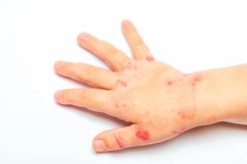
A healthy term infant born via normal vaginal delivery was noted at birth to have numerous small vesicles involving most of his face and upper chest. He was transferred to the neonatal ICU for suspicion of disseminated herpes simplex. On examination, the infant had small, 1- to 2-mm, superficial, clear vesicles that were confluent on the forehead, eyelids, nose, cheeks, neck, and upper back. A Tzanck test was negative for multinucleated giant cells.
A healthy term infant born via normal vaginal delivery was noted at birth to have numerous small vesicles involving most of his face and upper chest. He was transferred to the neonatal ICU for suspicion of disseminated herpes simplex. On examination, the infant had small, 1- to 2-mm, superficial, clear vesicles that were confluent on the forehead, eyelids, nose, cheeks, neck, and upper back. A Tzanck test was negative for multinucleated giant cells. Skin biopsy confirmed a diagnosis of miliaria crystallina.
Miliaria crystallina is a common disease of the eccrine sweat glands; it occurs in up to 5% of infants, most frequently around 1 week of life. Epidemiological studies do not show a race or sex predilection. Development of miliaria before the fourth day of life is unusual.1 The condition develops most often in warm or humid environments, including incubators, and after treatment with cholinergic or adrenergic medications.1,2 Additional risk factors include exposure to UV light, bacterial colonization of the skin with Staphylococcus epidermidis, and frequent sweating.2
Miliaria has been classified into various types on the basis of the level of obstruction of the eccrine gland; miliaria crystallina represents the most superficial occlusion-occlusion that occurs in the subcorneal and intracorneal epidermis.1
The lesions of miliaria crystallina are 1- to 2-mm noninflammatory clear vesicles on a clear base that easily rupture with minimal pressure. They appear on the forehead initially and may become generalized, with a predilection for areas covered by clothing and intertriginous areas.1,3 The clinical presentation is the result of eccrine duct obstruction leading to rupture, which causes sweat to become trapped in the skin. The cause of the obstruction is not understood, but bacteria may play a role in its pathogenesis.1 A scraping of a vesicle does not reveal inflammatory cells, and biopsy is not routinely indicated.3
The lesions of miliaria crystallina are generally asymptomatic, self-limited, and without complications. The vesicles rupture and desquamate without treatment several days after they appear. To facilitate regression of the rash, move affected infants to a cooler, less humid environment. Bathing patients in cool water can also hasten resolution of the rash.3 Although the rash clears within several days, anhidrosis takes 2 weeks to resolve; this coincides with the time necessary for eccrine gland turnover.2
Bacterial superinfection can occasionally complicate deeper variants of miliaria but is uncommon in miliaria crystallina.3 Miliaria crystallina is important to recognize because conditions with similar presentations are more serious; the latter include herpes simplex virus infection, staphylococcal scalded skin syndrome, congenital candidiasis, incontinentia pigmenti, and epidermolysis bullosa.1
References:
REFERENCE:
1.
Gan V, Hoang M. Generalized vesicular eruption in a newborn.
Pediatr Dermatol
. 2004;21:171-173.
2.
Wolff K, Goldsmith L, Katz S, Gilchrest B, eds.
Fitzpatrick's Dermatology in General Medicine
. 7th ed. New York: McGraw-Hill Companies; 2008:730.
3.
Harper J, Oranje A, Prose NS, eds.
Textbook of Pediatric Dermatology
. Oxford, UK: Wiley-Blackwell; 2006:63-65.
Articles in this issue
over 15 years ago
Atypical Dermatitis Herpetiformisover 15 years ago
Developing Pattern Recognition: The Key to Pediatric Dermatologyover 15 years ago
Drug Eruptions: The Benign-and the Life-Threateningover 15 years ago
Tips for Identifying and Treating Nail Disordersover 15 years ago
Black-Spot Poison Ivyover 15 years ago
Can You Distinguish Among These Diaper Dermatoses?over 15 years ago
Unexplained Bruising: Weighing the Pros and Cons of Possible Causesover 15 years ago
Is this a dermatophyte infection of the scalp?Newsletter
Access practical, evidence-based guidance to support better care for our youngest patients. Join our email list for the latest clinical updates.


