- January/February 2022
- Volume 39
- Issue 1
Generalized, eruptive lichen planus in a pediatric patient
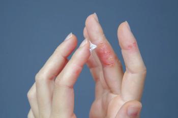
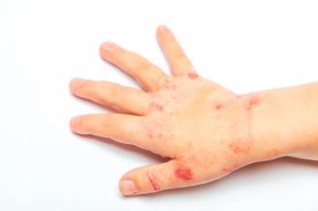
A healthy 14-year-old boy presented at our dermatology practice with acute onset of an intensely itchy rash that first appeared 2 months prior.
The case
A healthy 14-year-old boy presented at our dermatology practice with acute onset of an intensely itchy rash that first appeared 2 months prior (Figures 1 and 2). The patient had no family history of skin disease, was on no medications, and denied a history of preceding viral symptoms.
Diagnosis: Lichen planus
Clinical findings
The rash consisted of symmetrically and diffusely scattered violaceous to hyperpigmented papules with overlying silver scale coalescing into plaques. Affected areas included the entire trunk, extremities, and genitals. A punch biopsy demonstrated a lichenoid dermatitis consistent with lichen planus. In light of the presentation, the findings were consistent with pediatric eruptive generalized lichen planus (LP).
Etiology/epidemiology
LP is a pruritic, inflammatory dermatitis first described in 1869. The clinical appearance is classically described by the 6 P’s: purple, polygonal, planar, pruritic papules, and plaques. Additionally, a network of fine, reticular white lines called Wickham striae may be seen within the skin and mucosal lesions. Similar to psoriasis, The Koebner phenomenon (skin lesions appearing in areas of skin trauma, especially scratching and picking), which is also seen in psoriasis, is classically seen with LP.1 Several clinical subtypes, based on morphological pattern, have been described. Classic LP (also known as papular) is the most common subtype.2 LP variants include actinic, annular, atrophic, hypertrophic, inverse, bullous, and ulcerative.
LP is rarely seen in children, with just 1% to 4% of total cases diagnosed in the pediatric population.3 Children are more likely to present with oral mucosal involvement occurs more than over half of adult patients and may be the only site of disease, it is rare in children.4 Pruritis, a hallmark symptom of LP in adults, is frequently absent in children. Nail involvement, which can include nail thinning, pterygium (overgrowth of nail fold onto nail bed of the skin surrounding the nail and the skin under the nail plate), and subungual hyperkeratosis (thickened skin underlying the nail plate), is more common in adults compared with children.
The most common pattern in pediatric patients is the classic form.3,5 The second most common form in children is eruptive (16%),3 as seen in this patient. Many children have a polymorphic and heterogenous disease phenotype1; in these cases, LP is classified based on the most dominant pattern. Pediatric patients are more likely than adults to have a more severe disease course.3
A variety of drugs and dental materials have been implicated as inciting factors.4 Several autoimmune diseases have been linked to LP, including vitiligo, Hashimoto thyroiditis, myasthenia gravis, and alopecia areata.3 Other disease associations include liver disease (particularly hepatitis C and primary biliary cholangitis6) and thymoma. The aforementioned associations have been described in adult patients with LP. It is important to note, however, that these risk factors do not appear to apply to pediatric patients.
Several studies show a disproportionate number of cases of pediatric LP in the Indian subcontinent. Here, Black children may be more affected than other races.6
There is evidence that CD8+ T cells are strongly implicated in the pathogenesis of LP because they cause apoptosis (programmed cell death) of keratinocytes, but the true pathogenesis remains unknown. Diagnosis is typically confirmed by punch biopsy.
Differential diagnosis
The differential diagnosis for LP can be extensive, but 2 common considerations are lichenoid drug eruption (LDE) and psoriasis. A history of medications that can trigger an LDE, including antihypertensives, diuretics, and nonsteroidal anti-inflammatory drugs, is critical in differentiating it from LP.7
LP and psoriasis are both considered papulosquamous disorders. One key differentiator is the presence of Wickham striae in LP compared with overlying scale in psoriasis. LP can also remit without treatment after about 1 year, whereas psoriasis is generally a lifelong condition.
Treatment and management
There is no cure for LP, and treatment is aimed at managing symptoms. Topical corticosteroids are often used as a first-line option, and most patients show an excellent response.3 For treatment-resistant disease, systemic steroids and dapsone can be prescribed. Following resolution of the rash, postinflammatory hyperpigmentation (PIH) and cicatricial alopecia can be seen. Management of PIH includes topical hydroquinone and a retinoid.3
With treatment, pediatric LP generally carries a good prognosis. However, the eruptive pattern of LP can have severe, acute manifestations. In addition, long-term complications of LP involving the genitals can include scarring, urethral strictures, and urethral stenosis.8 Although there is no associated risk of skin cancer, LP with oral involvement has been linked to the development of oral squamous cell carcinoma.9 These potential complications underscore the need for prompt treatment.
Patient outcome
The patient started on triamcinolone 0.1% ointment twice daily for affected areas, hydrocortisone 2.5% ointment twice daily for the genital area, and oral hydroxyzine 25 mg nightly.
At follow-up 2 weeks later, the patient had mild improvement in pruritus and appearance of the rash. Fluocinonide 0.05% ointment twice daily was added for the most pruritic areas. At 8 weeks’ follow-up, pruritus had resolved and the rash had improved, with a few remaining foci of activity and residual PIH (Figure 2). Hydroxyzine and fluocinonide were stopped, and oral metronidazole 500 mg twice daily was added. A prior authorization for narrow band UV-B 3 times per month for 3 months was submitted.
At 12 weeks’ follow-up, metronidazole 500 mg twice weekly was continued for an additional 4 weeks, as was triamcinolone 0.1% ointment twice daily to the worst areas and hydrocortisone 2.5% ointment twice daily to the less involved areas.
References
1. Ott H, Frank J, Poblete-Gutiérrez P. Eruptive lichen planus in a child. Pediatr Dermatol. 2007;24(6):637-639. doi:10.1111/j.1525-1470.2007.00553.x
2. Gorouhi F, Davari P, Fazel N. Cutaneous and mucosal lichen planus: a comprehensive review of clinical subtypes, risk factors, diagnosis, and prognosis. Sci World J. 2014:742826. doi:10.1155/2014/742826
3. Pandhi D, Singal A, Bhattacharya SN. Lichen planus in childhood: a series of 316 patients. Pediatr Dermatol. 2014;31(1):59-67. doi:10.1111/pde.12155
4. Khandelwal V, Nayak PA, Nayak UA, Gupta A. Oral lichen planus in a young Indian child. BMJ Case Rep. 2013;2013:bcr2013010516. doi:10.1136/bcr-2013-010516
5. Nnoruka EN. Lichen planus in African children: a study of 13 patients. Pediatr Dermatol. 2007;24(5):495-498. doi:10.1111/j.1525-1470.2007.00501.x
6. Walton KE, Bowers EV, Drolet BA, Holland KE. Childhood lichen planus: demographics of a U.S. population. Pediatr Dermatol. 2010;27(1):34-38. doi:10.1111/j.1525-1470.2009.01072.x
7. Lichenoid drug eruption. DermNet NZ. Accessed August 29, 2020.
8. Machin SE, McConnell DT, Adams JD. Vaginal lichen planus: preservation of sexual function in severe disease. BMJ Case Rep. 2010;2010:bcr08.2009.2208. doi:10.1136/bcr.08.2009.2208
9. Silveira W da S, Bottezini EG, Linden MS, et al. Squamous cell carcinoma from oral lichen planus: a case report of a lesion with 28 years of evolution. J Korean Assoc Oral Maxillofac Surg. 2017;43(suppl 1):S14-S18. doi:10.5125/jkaoms.2017.43.s1.s14
Articles in this issue
almost 4 years ago
What might 2022 hold?almost 4 years ago
Severe hemorrhage from infantile hemangiomaalmost 4 years ago
Harmful excipients for pediatric patientsalmost 4 years ago
Crossover symptoms of COVID-19 and influenzaalmost 4 years ago
Eating habits of children during COVID-19about 4 years ago
Are you screening for autism when—and as often as—you should?about 4 years ago
Gestational diabetes can have lasting effects on child healthNewsletter
Access practical, evidence-based guidance to support better care for our youngest patients. Join our email list for the latest clinical updates.


