- Consultant for Pediatricians Vol 2 No 6
- Volume 2
- Issue 6
Staphylococcal Scalded Skin Syndrome in a 2-Year-Old Girl
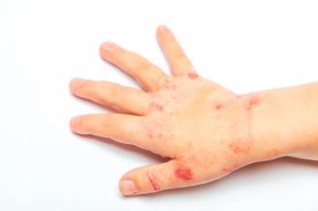
A toddler was brought to the emergency department with a 3-day history of a rash on her neck and irritability and fever (temperature, 38.3°C [101°F]) of 1 day's duration. The child's mother noticed a red, purulent bump on the girl's hand 2 days before the rash developed.
A toddler was brought to the emergency department with a 3-day history of a rash on her neck and irritability and fever (temperature, 38.3°C [101°F]) of 1 day's duration. The child's mother noticed a red, purulent bump on the girl's hand 2 days before the rash developed.
The slightly ill-appearing but well-developed 2-year-old had periorbital and perioral erythema with desquamation. Confluent areas of erythema and desquamation with scattered areas of crust and ulceration also were apparent on her neck. The oral mucosa and conjunctiva were clear. The lesion on the dorsum of the girl's hand was a 7-mm, faintly erythematous papule with a central crust and no purulent discharge.
Laboratory findings revealed mild leukocytosis: white blood cell count of 10,100/µL, with 58% neutrophils, 30% lymphocytes, 4% monocytes, and 3% eosinophils. Clinical chemistry and urinalysis results were within normal limits. Throat, blood, and conjunctival cultures were negative.
A diagnosis of staphylococcal scalded skin syndrome (SSSS) was made; the pyoderma on the hand was the source of the infection.
SSSS is caused by a strain of Staphylococcus aureus that produces an exfoliative toxin which causes cleavage of the upper layers of the epidermis, bullae formation and, ultimately, sloughing of the skin. The bacteria are not found in the lesions but at a distant focus; the conjunctiva, nasopharynx, skin, and stools are common sites.
SSSS is seen primarily in children younger than 5 years. Characteristic early signs of the disease are malaise; fever; irritability; and a tender, macular erythema that is most prominent in periorificial and intertriginous areas but may become generalized. The exfoliative phase begins with periorificial exudation and crusting that spreads peripherally. The skin develops a wrinkled appearance and can be removed with light stroking. With prompt and proper antibiotic treatment, healing without scarring takes place in 2 weeks.
Because of their nearly identical clinical appearance, SSSS must be differentiated from toxic epidermal necrolysis (TEN). TEN, which chiefly affects adults, is a reaction to a drug-most commonly an anticonvulsant or a sulfonamide. Mucous membrane involvement is common in TEN; the cleavage point is at the dermal-epidermal junction, whereas the cleavage point in SSSS occurs in the stratum corneum. If the diagnosis is uncertain, a frozen section of a blister roof can be examined.
Treatment of SSSS includes antibiotics effective against S aureus and local wound care with bland emollients, such as hydrophilic petrolatum. Bacitracin, neomycin, and similar agents need to be used cautiously, since they may cause allergic contact dermatitis. A silver sulfadiazine preparation is useful but requires frequent applications. Mupirocin ointment provides excellent S aureus coverage and is a very good emollient.
This youngster was treated with intravenous nafcillin and topical hydrophilic petrolatum ointment. The 2-week follow-up examination was significant only for postinflammatory hyperpigmentation over the involved areas.
FOR MORE INFORMATION:
- Amon RB, Dimond RL. Toxic epidermal necrolysis: rapid differentiation between staphylococcal and drug-induced disease. Arch Dermatol. 1975;111:1433-1437.
- Borchers SL, Gomez EC, Isseroff RR. Generalized staphylococcal scalded skin syndrome in an anephric boy undergoing hemodialysis. Arch Dermatol. 1984;120:912-918.
- Elias PM, Fritsch P, Epstein EH. Staphylococcal scalded skin syndrome: clinical features, pathogenesis, and recent microbiological and biochemical development. Arch Dermatol. 1977;113:207-219.
- Freedberg IM, Eisen AZ, Wolff K, et al, eds. Fitzpatrick's Dermatology in General Medicine. 5th ed. New York: McGraw-Hill; 1999.
- Rothe MJ, Grant-Kels JM. Dermatologic emergencies: part 5: drug-induced Stevens-Johnson syndrome and toxic epidermal necrolysis. Consultant. 1998;38:889-895.
Articles in this issue
over 20 years ago
Bullous Impetigo in a 3-Year-Old Girlover 22 years ago
Herpangina in a 17-Year-Old Boyover 22 years ago
Erysipelasover 22 years ago
Complications of ChickenpoxNewsletter
Access practical, evidence-based guidance to support better care for our youngest patients. Join our email list for the latest clinical updates.


