Consultant for Pediatricians Vol 9 No 6

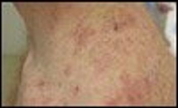
An 18-year-old boy presented with a several-month history of an intermittent, very pruritic rash on his back that did not improve with topical corticosteroids. Physical examination revealed grouped erythematous papules with a few scattered small vesicles on his posterior neck and bilateral posterior shoulders at the location where his backpack frequently rubbed.

A healthy term infant born via normal vaginal delivery was noted at birth to have numerous small vesicles involving most of his face and upper chest. He was transferred to the neonatal ICU for suspicion of disseminated herpes simplex. On examination, the infant had small, 1- to 2-mm, superficial, clear vesicles that were confluent on the forehead, eyelids, nose, cheeks, neck, and upper back. A Tzanck test was negative for multinucleated giant cells.

After completing training in pediatrics, dermatology, and pediatric dermatology, I am convinced that the art of medicine, especially as practiced in the field of pediatric dermatology, consists largely of an ability to use pattern recognition to separate the usual from the rare.

“Drug rash” is a common pediatric complaint in both inpatient and outpatient settings. This term, however, denotes a clinical category and is not a precise diagnosis. Proper identification and classification of drug eruptions in children are important for determining the possibility of-and preventing progression to-internal involvement. Accurate identification is also important so that patients and their parents can be counseled to avoid future problematic drug exposures.

Disorders of children’s fingernails and toenails can often be difficult to diagnose or treat. Here I provide tips on identifying and treating some of the nail disorders seen in pediatric practice.

A healthy 9-year-old girl presented with pruritic, darkly colored papules and linear lesions on the left side of her face. She had recently been on a hiking trip with her family. Examination revealed erythematous papules and linear streaks with an overlying black substance that resembled black lacquer paint.

A 1-year-old boy presented with a 10-day history of a nonpruritic rash that had persisted and spread despite treatment with a topical corticosteroid. Mother reported that he was febrile at the onset of the eruption; he was given over-the-counter antipyretics. On day 3, his pediatrician evaluated his condition and prescribed amoxicillin for his fever and hydrocortisone cream for his atopic dermatitis. Over the next several days, the fever subsided; however, the rash, which had started on the child’s right hand, persisted and spread to his face and elsewhere.

A 3-month-old boy was brought by his mother to his busy primary care physician’s office for follow-up of bronchiolitis when numerous bruises were noted. The mother said that the infant had a 1-week history of unexplained bruising, petechiae, and irritability. The child was referred to the local emergency department (ED) because of concern for nonaccidental trauma.

Invasive pneumococcal disease (IPD) in children can cause serious illness-including meningitis, pneumonia, and bacteremia-and death. Fortunately for children, their families, and their pediatricians, the incidence of IPD in children younger than 5 years has dropped significantly following the widespread adoption of the pneumococcal conjugate vaccine (PCV).

The mother of this 7-year-old girl originally thought these peculiar scales in her daughter’s scalp and hair were nits, since there had recently been an outbreak of head lice at the child’s school. However, she was not able to find any lice, and the scales resisted removal with mineral oil and a “nit comb.”

Four hours after a 12-year-old boy was stung by a honeybee on his right middle finger, he noticed localized hand swelling, erythema, and tenderness. The following morning, his entire hand was swollen and erythematous, with contiguous erythema on the medial aspect of his forearm and arm. Although the arm was tender to palpation, he was afebrile and felt well. Because of an initial concern for cellulitis or lymphangitis, he was given intravenous antibiotics and antihistamines and was admitted overnight for observation.

During a well adolescent visit, an obese 17-year-old boy complained of left knee pain of 4 years’ duration. The pain was worse at night. He was able to ambulate. He associated the pain with a left tibial fracture he sustained after falling off a bicycle 4 years earlier; he denied recent trauma.

A hemangioma can be concerning to parents, who want to know the prognosis for the lesion as their infant grows. Here, clues to help you identify those that will rapidly involute, those that will grow for a while and then involute over a period of years, and those that without treatment will remain unchanged. Also, which hemangiomas warrant referral.