- Consultant for Pediatricians Vol 4 No 5
- Volume 4
- Issue 5
Case In Point: Toxic Shock Syndrome
A 12-year-old boy was brought by ambulance to the emergency department (ED) with fever and shaking of 3 days' duration. He was accompanied by his mother. The boy had spent the weekend at his father's home when he began to feel sick. Since returning to his mother's house, he has been lethargic and has had one episode of vomiting.
Fever is one of the most common symptoms for which children are brought to the emergency department. In most cases, fever is attributed to viral infections; in many cases, causes such as otitis media, pneumonia, and urinary tract infection are identified. Occasionally, however, one comes across an unusual case of fever accompanied by a sudden drop in blood pressure. A systematic approach to diagnosis and management is crucial in preventing morbidity and mortality.
THE CASE
A 12-year-old boy was brought by ambulance to the emergency department (ED) with fever and shaking of 3 days' duration. He was accompanied by his mother. The boy had spent the weekend at his father's home when he began to feel sick. Since returning to his mother's house, he has been lethargic and has had one episode of vomiting.
A year earlier, the patient had experienced a generalized seizure. A CT scan taken at that time showed an arachnoid cyst in the brain. The boy had been taking phenytoin, but his mother had discontinued the medication (without the physician's knowledge) a few weeks before the current ED visit because he had no new seizures and was "doing fine." He had also been taking methylphenidate for hyperactivity for the past year.
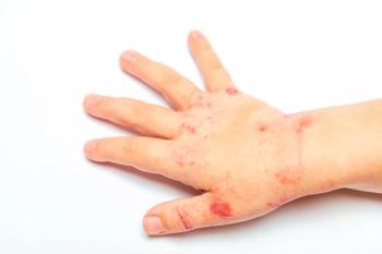
In the ED, the boy's temperature was 40.2ºC (104.3ºF); he had not received any medication for fever at home. His heart rate was 146 beats per minute; respiratory rate, 22 breaths per minute; and blood pressure, 132/49 mm Hg. He was unable to sit up or stand. He appeared flushed, especially over his palms and soles; the erythema blanched with pressure. The sclera were icteric. There was a small, 0.5-cm nontender pustule on his left knee. The liver and spleen were not enlarged.
The boy was lethargic and responded to questions in monosyllables. Meningeal signs were absent. By the time the examination was complete, his blood pressure had dropped to 90/58 mm Hg. He received a bolus of normal saline, and ceftriaxone therapy was started after blood and urine samples were obtained for culture. A sample of the content of the pustule on his knee was also sent for culture. The boy was given dopamine to maintain his blood pressure and was admitted to the pediatric ICU (PICU).
In the PICU, the patient's white blood cell (WBC) count was 17,000/µg, with 15% bands, 71% neutrophils, 6% lymphocytes, and 8% monocytes. His hemoglobin was 13.2 g/dL; platelet count, 118,000/µg; reticulocyte count, 1.2%. Serum sodium level was 131 mEq/L; potassium, 4.3 mEq/L; chloride, 98 mEq/L; bicarbonate, 19 mEq/L; blood urea nitrogen, 1 mg/dL; and creatinine, 0.8 mg/dL. Urinalysis revealed the following: pH, 5.5; 2+ bilirubin; 1+ ketone; 2+ protein; 4 mg/dL urobilinogen; 10 to 20 WBCs; and 3+ bacteria. Serum bilirubin level was elevated at 5 mg/dL (direct fraction, 1.9 mg/dL). Levels of aspartate aminotransferase, alkaline aminotransferase, alkaline phosphatase, and creatine kinase were normal, as were prothrombin time and partial thromboplastin time. The d-dimer level in serum was mildly elevated at 3.61 mg/L. Urine was negative for myoglobin. Clindamycin was added to his antibiotic therapy.
Over the next 24 hours, the patient's blood pressure remained stable and the dopamine drip was tapered gradually. Blood and urine cultures at 24 hours showed no growth. The CT scan demonstrated a 3-cm right temporal arachnoid cyst without any acute changes. The culture from the pustule on the boy's knee grew coagulase-positive Staphylococcus. Staphylococcus aureus also grew from the nasopharyngeal swab. Staphylococcal toxic shock syndrome (TSS) was diagnosed.
The boy improved rapidly after 24 hours, and blood pressure support was withdrawn by 48 hours of admission. The erythema disappeared in 4 days, and he was sent home 5 days after admission on a 10-day course of oral clindamycin. The patient was doing well at home 10 days later and reported some generalized desquamation.
ABOUT TSS
TSS is uncommon in children. Todd and colleagues1 initially described this entity in 1978 in 7 children between 8 and 17 years of age. TSS was later recognized to be more common in menstruating girls and women who used tampons. The incidence of menstrual-related TSS has declined since highly absorbent tampons were withdrawn from the market in the 1980s. Currently, nonmenstrual TSS accounts for most cases.2 The recognition and early management of TSS has reduced associated mortality.
TSS is caused by massive amounts of inflammatory cytokines produced as a result of stimulation of T lymphocytes by staphylococcal superantigen--the TSS toxin-1 (TSST-1). This toxin has a 3-dimensional structure that allows it to interact with relatively invariant regions of major histocompatibility complex class II molecules on the surface of antigen-presenting cells and with certain variable regions of the T-cell receptor chain. Unlike a typical antigen that may activate 1 in 10,000 T cells, superantigens may activate from 20% to 50% of T cells. The consequence of these interactions is the exaggerated release of bioactive cytokines, which are responsible for the clinical signs of illness associated with these toxins.3
DIAGNOSIS
The diagnosis of TSS is mainly clinical. The CDC case definition requires the presence of 5 criteria (Table).4 Nonmenstrual TSS may present differently from menstrual TSS in that the onset of symptoms may be delayed after the precipitating injury or event; also, there are more frequent CNS manifestations, less frequent musculoskeletal involvement, and more severe anemia.5 TSS is associated with a 2% to 5% mortality rate: early diagnosis is therefore crucial. A high index of suspicion--especially in patients who present with fever and shock without obvious cause--is particularly important. As in this patient's case, the offending staphylococcal lesion can be small and may be unnoticed by the patient.
The problems we encountered during diagnosis in our patient included the appearance of jaundice and hemoglobinuria, which swayed our diagnosis toward other hemolytic disorders. While hyperbiliru- binemia is a known finding in staphylococcal TSS, the cause in the absence of raised liver enzyme levels remains uncertain. This could be a result of hepatic underperfusion resulting from shock or a direct effect of staphylococcal toxin.6
The immediate need for patients with TSS is aggressive fluid therapy and management of respiratory and/or cardiac failure. Initial empiric antibiotic therapy is with a combination of a b-lactamase-resistant antimicrobial agent and a protein synthesis-inhibiting agent, such as clindamycin. Inhibition of protein synthesis leads to diminished toxin synthesis. Antimicrobial therapy should be continued for a minimum of 10 days to ensure eradication of the organism, because recurrences are known to occur.7
In addition, any abscesses should be drained. Consider intravenous immunoglobulin (IVIG) therapy if the area of infection cannot be drained; also consider this treatment for patients who respond poorly to standard treatment.3,4 Standard IVIG preparations contain neutralizing antibodies against the superantigens responsible for the clinical manifestations.
BOTTOM LINE
The mortality associated with TSS is high if proper treatment is not instituted immediately. Therefore, in addition to managing the patient's airway, breathing, and circulation, look carefully for any infected lesions that suggest TSS. As demonstrated in this case, early and appropriate management can result in a rapid recovery.
References:
REFERENCES:
1. Todd J, Fishaut M, Kapral F, Welch T.Toxic-shock syndrome associated with phage-group-I staphylococci.
Lancet.
1978;2:1116-1118.
2. Gaventa S, Reingold A, Hightower AW, et al. Active surveillance for toxic shock syndrome in the United States, 1986.
Rev Infect Dis.
1989;11(suppl 1): S28-S34.
3. Schlievert PM. Use of intravenous immunoglobulin in the treatment of staphylococcal and streptococcal toxic shock syndrome and related illnesses.
J Allergy Clin Immunol.
2001;108(4 suppl):S107-S110.
4. American Academy of Pediatrics. Toxic shock syndrome. In: Pickering LK, ed.
Red Book
:
2003 Report of the Committee on Infectious Diseases.
26th ed. Elk Grove Village, Ill: American Academy of Pediatrics; 2003:624-630.
5. Kain KC, Schulzer M, Chow AW. Clinical spectrum of nonmenstrual toxic shock syndrome (TSS): comparison with menstrual TSS by multivariate discriminant analyses.
Clin Infect Dis.
1993;16: 100-106.
6. Gourley GR, Chesney PJ, Davis JP, Odell GB. Acute cholestasis in patients with toxic-shock syndrome.
Gastroenterology.
1981;81:928-931.
7. Andrews MM, Parent EM, Barry M, Parsonnet J. Recurrent nonmenstrual toxic shock syndrome: clinical manifestations, diagnosis, and treatment.
Clin Infect Dis.
2001;32:1470-1479.
Articles in this issue
over 20 years ago
Aniridiaover 20 years ago
Photoclinic: Herpes Simplex Virus Infectionover 20 years ago
Pediatric Urology Clinics: Dysuria in an Elementary School-Aged Boyover 20 years ago
Spitz Nevus and Granuloma Annulareover 20 years ago
Photoclinic: Winged Scapulaover 20 years ago
Photo Essay: Childhood Alopeciaover 20 years ago
"Club" Drugs 101over 20 years ago
Asthma Update: Pearls You May Have Missedover 20 years ago
Duodenal Atresia in a Neonateover 20 years ago
Recurrent Strep Throat: How Best to TreatNewsletter
Access practical, evidence-based guidance to support better care for our youngest patients. Join our email list for the latest clinical updates.


