- Consultant for Pediatricians Vol 23 No 5
- Volume 23
- Issue 5
Psoriasis in an 11-Month-Old Infant
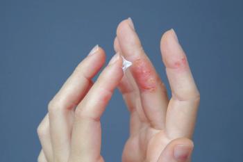
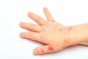
An otherwise healthy 11-month-old infant hadhad an intermittent, nonpruritic rash for mostof his life. The lesions recurred mainly onthe extremities and trunk without a particulartrigger. Applications of 1% hydrocortisonecream were only partially beneficial. The joints and nailswere not affected. The patient’s maternal grandfather hadsevere psoriasis.
An otherwise healthy 11-month-old infant had had an intermittent, nonpruritic rash for most of his life. The lesions recurred mainly on the extremities and trunk without a particular trigger. Applications of 1% hydrocortisone cream were only partially beneficial. The joints and nails were not affected. The patient's maternal grandfather had severe psoriasis.
Linda S. Nield, MD, of Morgantown, WVa, and Deepak M. Kamat, MD, PhD, of Detroit write that erythematous plaques with thick, silvery scales are characteristic of psoriasis. The plaque type is the most commonly observed variant in children, followed by guttate psoriasis and juvenile psoriatic arthritis.1 The head (especially the scalp), extremities, trunk, and diaper area are most frequently affected. The plaques may be pruritic but can be asymptomatic, as they were in this patient. Nail changes, such as pitting of the nail surface, are also a typical manifestation of psoriasis.
Scaly patches and plaques can occur with many skin disorders (ie, atopic dermatitis, dermatophyte infections, pityriasis rosea, and lichen planus). Collagen-vascular diseases, such as lupus and dermatomyositis, are associated with scaly plaques and joint disease in affected children. Signs of pharyngitis or perineal infection with group A streptococci (GAS) may be evident in children with guttate psoriasis; screening for such infection is necessary in these children. Joint abnormalities are present in children with juvenile psoriatic arthritis.
Characteristic clinical findings and a family history are adequate to make the diagnosis. The Köbner phenomenon, or isomorphic response--in which linear psoriatic lesions develop at a site of external trigger--is a useful diagnostic clue. Histologic examination of a lesion shows an inflammatory reaction associated with epidermal proliferation.
Both genetic and environmental factors play a role in the development of psoriasis. More than two thirds of children with psoriasis have a family history.2,3 Environmental triggers, such as physical injury or infection, can precipitate the disease.
Parental and child education about proper skin care is essential. A dermatologist should be consulted and actively involved in the management of psoriasis. Treatment is individualized and determined by the patient's age, disease severity, and response to first-line topical therapy. Emollients, corticosteroids, coal tar, dithranol, and calcipotriol are effective.4 UV-B phototherapy can be combined with topical agents.
Nonresponsive and severe disease may warrant systemic treatment. Psoralens plus UV-A phototherapy is an option. If retinoids, methotrexate, and cyclosporine are necessary, the safety concerns associated with the use of these medications in children must be kept in mind.4 Patients with guttate psoriasis who have documented infection with GAS require appropriate antibiotics.
Articles in this issue
about 20 years ago
Musculoskeletal Clinics: 12-Year-Old With Knee Pain From Kickball Injuryabout 20 years ago
Pediatric Urology Clinics: Red Urine in an 8-Year-Old Boyabout 20 years ago
Photoclinic: Swallowed Beadsabout 20 years ago
Infected Cystic Hygromaabout 20 years ago
Necrobiosis Lipoidica in an Adolescentabout 20 years ago
Case In Point: Infantile Hypertrophic Pyloric Stenosisabout 20 years ago
Halo Nevus and Nevus Spilusabout 20 years ago
Photoclinic: Imperforate Anus With Anocutaneous FistulaNewsletter
Access practical, evidence-based guidance to support better care for our youngest patients. Join our email list for the latest clinical updates.


