- Consultant for Pediatricians Vol 5 No 4
- Volume 5
- Issue 4
Pityriasis Rosea in a 7-Year-Old Girl
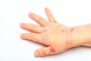
Seven-year-old girl with generalized rash that started as a single isolated oval lesion on the lower abdomen. Six days later, diffuse papulosquamous lesions appeared mainly on the trunk, sparing the scalp, face, and extremities. Intense itching despite 3 days of diphenhydramine therapy.
HISTORY
Seven-year-old girl with generalized rash that started as a single isolated oval lesion on the lower abdomen (A). Six days later, diffuse papulosquamous lesions appeared mainly on the trunk (B), sparing the scalp, face, and extremities. Intense itching despite 3 days of diphenhydramine therapy.
Patient otherwise healthy. No recent history of upper respiratory tract infection.
PHYSICAL EXAMINATION
Patient is afebrile; appears healthy. Initial oval lesion seen as a sharply defined area of dermatitis with a pinkish center and a slightly elevated scaly border. Subsequent lesions resembled the initial solitary lesion but were smaller. Lesions on the back (C) distributed in a Christmas tree pattern following skin cleavage lines. No other remarkable physical findings.
Pityriasis rosea (PR) is a self-limited inflammatory skin condition. The name is derived from Greek (pityriasis = scaly) and Latin (rosea = pink). It is characterized in up to 50% of cases by the initial appearance of a herald patch,1-4 a solitary oval scaly lesion that slowly expands, sometimes to several centimeters in diameter.
Secondary lesions appear approximately 2 to 21 days later. They continue to erupt in crops over the next 10 to 21 days and may persist for 4 to 10 weeks. The round to oval, 5- to 10-mm lesions have an elevated scaly border; they involve the trunk and, less commonly, the extremities. These lesions, when seen on the back, are distributed in a Christmas tree pattern following cleavage lines (Langer lines). The palms and soles are usually spared.
Pruritus affects approximately 25% of patients.2,3,5 There are no other systemic symptoms during the rash phase of PR. In dark-skinned persons, post-inflammatory hyperpigmentation or hypopigmentation may be seen after the lesions resolve. During the post-inflammatory phase, hypopigmentation may mimic pityriasis alba or tinea versicolor.
Some epidemiologic features (seasonal variation, clustering in communities) suggest that PR may be an infectious disease. At present, however, no causative agent has been identified. WHAT'S YOUR DIAGNOSIS?
DIAGNOSIS AND THERAPY
The diagnosis of PR is usually clinical. The characteristic physical findings usually lead to the correct diagnosis. However, when only the herald patch is present, tinea corporis and nummular eczema should be considered. Guttate psoriasis usually manifests with silvery scales that are thicker than those seen in PR.
If the diagnosis is uncertain, or if the patient is sexually active and his or her palms and soles are involved, consider secondary syphilis.
Treatment is usually symptomatic and is mainly directed toward relieving pruritus. Patient education and reassurance are the most effective form of therapy. The parents and the patient need reassurance that the rash will resolve spontaneously within 4 to 10 weeks.
References:
REFERENCES:
1.
Tay YK, Goh CL. One-year review of pityriasis rosea at the National Skin Centre, Singapore.
Ann Acad Med Singapore.
1999;28:829-831.
2.
Bjornberg A, Hellgren L. Pityriasis rosea. A statistical, clinical, and laboratory investigation of 826 patients and matched healthy controls.
Acta Derm Venereol.
1962;42(suppl 50):1-68.
3.
Allen RA, Janniger CK, Schwartz RA. Pityriasis rosea.
Cutis.
1995;56:198-202.
4.
Jacyk WK. Pityriasis rosea in Nigerians.
Int J Dermatol.
1980;19:397-399.
5.
Gonzalez LM, Allen R, Janniger CK, Schwartz RA. Pityriasis rosea: an important papulosquamous disorder.
Int J Dermatol.
2005;44:757-764.
Articles in this issue
almost 20 years ago
Day-Old Boy With Respiratory Distress After Complicated Deliveryalmost 20 years ago
Genetic Disorders: Child With Facial Anomalies and Developmental Delayalmost 20 years ago
A 7-Year-Old Boy With Blistering Skinalmost 20 years ago
Guest Commentary: If I Have Herpes, Can I Still Have Children? . . .almost 20 years ago
Neonatal Acne in a 3-Week-Old Boyalmost 20 years ago
Photoclinic: Inflamed Keratosis Pilarisalmost 20 years ago
Case in Point: A Young Girl With Cafe au Lait SpotsNewsletter
Access practical, evidence-based guidance to support better care for our youngest patients. Join our email list for the latest clinical updates.


