
IP is a rare X-linked dominant disorder. About 700 to 1000 cases have been reported worldwide (about 1 in 50,000 live births); white infants are most commonly affected. In a review of 653 patients, more than half had a family history of the condition.1 Our patient's mother was also affected. IP usually appears within the first 2 weeks of life. The severity and expression of the disorder are highly variable, even among patients within the same family.1,3 The condition is characterized by anomalies of the organs and tissues derived from the ectoderm and mesoderm and may affect the skin, hair, nails, teeth, eyes, and CNS1,2:
Dermatology
Latest News
Advertisement
Advertisement
This self-limited unilateral dermatitis of unknown cause usually affects preschool children.1-3 Girls are 2 to 3 times more frequently affected than boys.4 The eruption consists of flat-topped pink or flesh-colored papules with a fine scale that form a linear band of less than 1.2 cm in width. It often follows the lines of Blaschko and may extend the entire length of an extremity.
ABSTRACT: Children who present withrash and fever can be roughly dividedinto 3 groups: the first group includesthose with features of serious illnesswho require immediate intervention. Thesecond and third groups include thosewith clearly recognizable viral syndromes,and those with early or undifferentiatedrash. Here the focus is on those childrenin group 1 who have petechiae or purpura.The morphology of lesions amongchildren with symptoms of serious illnessoffers clues to the underlying cause.For example, petechiae may herald suchlife-threatening disorders as meningococcemia,Rocky Mountain spotted fever,and hemolytic uremic syndrome.
Prominent ear deformity is relatively common; this defect- inherited as an autosomal dominant trait-affects approximately 5% of white children.1 As such, surgical correction is a common operation performed by plastic surgeons.
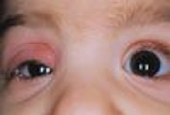
This 20-month-old child has had a localized red mass on the right eyelid that had been present for a few weeks.
Seventeen-month-old Hispanic boy with 7-month history of a swelling on his back. Lesion first looked like a small "scar," then gradually grew over next few months. Lesion appeared to blister, with subsequent discharge of clear fluid. No symptoms other than intermittent pruritus at lesion site. Patient not taking any medications.
Diagnosis and treatment of congenital midline cervical cleft.
This girl was brought for evaluation of these "bumps" around her eyes. Her parents are concerned that the lesions will interfere with her vision.
A few days before presentation, the mother noted some "bumps" that had developed behind the child's right ear. The child was brought to the emergency department for evaluation.
The "Baghdad boil," as it is known in Iraq, usually begins as a papule at the site of a sandfly bite. This progresses to a nodule and then an ulcer, which eventually crusts over. The ulcer is typically painless, unless infected. The female sandfly is a common vector of transmission of cutaneous leishmaniasis. Most cases are caused by Leishmania major in dry desert areas and Leishmania tropica in urban areas. Tissue biopsy remains the gold standard diagnostic test for the disease.2,4,5
The patient had no recent fevers, cough, or weight loss. His medical history was notable for chronic thrombocytopenia and reactive arthritis, for which he had been hospitalized. His maternal grandmother had systemic lupus erythematosus; his mother had died of congestive heart failure and emphysema.
17-Year-old female with a salmon-pink oval patch on her back. Three days after the patch appeared, a generalized eruption developed on her back. Eruption slightly pruritic. No antecedent upper respiratory tract infection. No prodromal symptoms.
Multiple tender flaccid bullae (about 2 mm to 2 cm) were present on the perineum. There was no associated discharge. The ruptured blisters had marked erythema in the center and a scaly rim at the periphery. The Nikolsky sign could not be elicited. The infant was afebrile. The remainder of the examination findings were normal.
Abbott's Humira (adalimumab) had been approved by the food and Drug Administration for its sixth indication, juvenile rheumatoid arthritis...
On morning rounds in the well-baby nursery, a nurse brings your attention to a 1-day-old girl who is having trouble latching onto the breast. You examine the child and note the subtle anomalies shown in Figure 1 along with a pronounced head lag and a systolic heart murmur.
As a practicing pediatrician in East Hampton, NY, where Lyme disease is endemic, I read with interest the recently published case by Riva Kamat, MD, involving a girl with Lyme meningitis who underwent a lumbar puncture.1
I always find it difficult to speak with pediatricians about diaper rashes. Pediatricians look after many more children with rashes in the diaper area than I do--and all have their own secret ways to treat these children.
For 3 weeks, a 3-year-old African American boy had a mildly pruritic rash on his buttocks, lower extremities, upper thighs, and soles. The patient was initially seen at an urgent care center, where he was given amoxicillin for suspected scarlet fever. A week later, he presented to the emergency department and was treated with griseofulvin for tinea corporis. A skin culture did not grow fungus.
During spring vacation, a previously healthy 4-year-old girl visited western Nebraska, where she and her family spent time along a river bank in a wooded area. After 4 days, her mother noticed 3 engorged ticks embedded in the child's scalp. The ticks were immediately removed and burned. The child also had a marble-sized swelling on the right side of her neck. Over the next few days, the child had temperatures that spiked to 39.4C (103F), with chills, generalized malaise, and weakness. There was no history of cough, myalgias, or headache.
Most physicians in this area of New York do not do spinal taps on patients with headaches and an erythema chronicum migrans rash unless they have meningismus. Seventh cranial nerve palsy alone is not an indication for a spinal tap.
This skin abnormality is cutis marmorata-a physiological dilatation of capillaries and venules of the trunk and extremities in infants and young children caused by exposure to cold. The discoloration fades with warming, as was the case with this baby. The condition is seen especially when subcutaneous fat is decreased.
The photos presented this month reveal disease entities I have seen that did not respond to conventional therapy and that became resistant "diaper rashes." You may have seen some of these "bottoms" in previous issues of Consultant For Pediatricians. Next to each photograph, I have given several clues to the diagnosis. See if you can match these clues with the diagnostic choices listed below. You can check to see whether your diagnostic choices are correct on page 61.
On morning rounds in the well-baby nursery, a nurse brings your attention to a 1-day-old girl who is having trouble latching onto the breast. You examine the child and note the subtle anomalies shown in Figure 1 along with a pronounced head lag and a systolic heart murmur.
The patient denied use of new skin products, detergents, or medications. He had no pets. There was no history of recent travel, and the patient was not aware of any arthropod bites. None of his family members had a similar rash. The patient was sexually active and had had 3 partners in the past 2 years; he said he always used condoms. His history was otherwise unremarkable, as were physical findings.
The child's mother recalled that the rash started as a single patch on his lower back and that the child had an upper respiratory tract infection 4 weeks earlier.
Advertisement
Advertisement
Trending on Contemporary Pediatrics
1
FDA approves acetaminophen-naproxen sodium combination tablet for ages 12 and older
2
US measles cases surpass 2025 total, hit highest level since 1991
3
Pooled phase 3 data show early, consistent tapinarof cream responses across pediatric age groups in AD
4
Prenatal therapy improves outcomes in fetal renal failure
5