
Dermatology

Latest News


A 3-week-old boy was referred for evaluation of suspected herpes simplex virus (HSV) infection in the inguinal and pelvic regions. The rash had reportedly worsened since its appearance 2 days earlier and was associated with a foul smell.

A 3-week-old boy was referred for evaluation of suspected herpes simplex virus (HSV) infection in the inguinal and pelvic regions. The rash had reportedly worsened since its appearance 2 days earlier and was associated with a foul smell.

Infantile seborrhea has many of the features of the other papulosquamous conditions listed, but the diagnosis is most commonly clinically evident on examination alone. If you are unsure, a KOH preparation will quickly sort out the dermatophytes. Also, the lack of itch makes atopic dermatitis very unlikely.

I believe that there is an entity termed "twenty-nail dystrophy" that is a benign and idiopathic disorder. The nail plates have a distinct appearance and all 20 nails are dystrophic. The nail plates are lusterless and their surface appears to have been sandpapered in a longitudinal direction. The plates are thin and rough with accentuated longitudinal ridges and the cuticles appear to be "roughed up" and thickened. The free ends of the plates are brittle and rough. The nail plates do not appear to be growing.

With the banning of peanut butter and jelly from someschool cafeterias, peanut allergies have become a populartopic in the media and the public. Discussions ofteninclude references to an increasing prevalence ofallergies, as well as to an earlier emergence of thoseallergies in children.

A 23-month-old Hispanic boy was brought to an emergency department (ED) with ear pain and fever. The family had no other expressed concerns. Physical examination revealed inflammation of 1 tympanic membrane. The child had a temperature of 38.4°C (101.2°F).

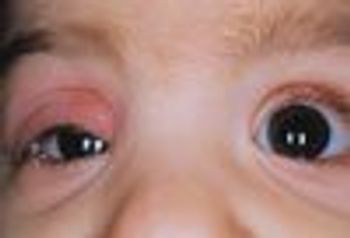
ABSTRACT: Routine screening for eye disease at all well-child visits should begin in the newborn period. Prompt ophthalmological referral of patients with strabismus or any suspected eye disease is essential to determine the underlying cause, optimize treatment, and preserve binocular vision.

An otherwise healthy female infant presented at birth with a lesion on the left side of the face that involved the frontotemporal scalp, periocular area, nose, and upper lip (A). She was born at term via spontaneous vaginal delivery after an uncomplicated gestation. The initial clinical impression was that of a port-wine stain associated with Sturge-Weber syndrome.

Photo A shows a red, slightly raised, 1 3 2-cm lesion on the left buttock of a 3-month-old boy. The lesion had been present since birth and had enlarged as the child grew. Photo B shows a similar lesion in the vulva of a 2-month-old girl that also had been present since birth; the mother stated that it had not enlarged.

The parents of this 12-month-old girl are concerned that their daughter “caught” psoriasis at her day-care center. The rash had been present for at least 6 months, after which it cleared significantly, only to return weeks later. The child does not seem to be bothered by it.

Ring around the nevus

An adolescent presents with several moles surrounded by white rings on his back.

This baby was born with an ulnar supernumerary digit on the left hand. This common congenital anomaly can be an isolated malformation or associated with other syndromes at birth.1-4 Although the true prevalence is unknown (because most hospitals do not report cases), the condition appears to be more common among males and African American infants.1-3 In African American infants, postaxial polydactyly is generally the result of autosomal dominant transmission. In white infants, the condition is frequently syndromic and is linked with autosomal recessive transmission.1

One week after vaccination with diphtheria, tetanus, and acellular pertussis/inactivated poliovirus/hepatitis B, Haemophilus influenzae type b, pneumococcal conjugate, and rotavirus, this 2Z\x-month-old infant presented with a vesicular rash. No other children in the home had a rash. The infant's primary caregiver was the grandmother, who had shingles 2 weeks earlier.

After experiencing frequent headaches, this 10-year-old girl awoke with a "funny smile" involving left-sided facial paresis. She was evaluated in an emergency department and a complete blood cell count, Lyme IgM and IgG screen, and CT scans of the head and sinuses were ordered.

ABSTRACT: Chronic recurrent multifocal osteomyelitis (CRMO) is an inflammatory bone disease that occurs primarily in childhood. The clinical picture often is confused with bacterial osteomyelitis. Awareness of CRMO as a clinical entity helps avoid diagnosis and treatment delays. Our patient, an 8-year-old girl, presented with acute left hip pain. One month after presentation, a lytic lesion was seen on plain radiographs; biopsy revealed nonspecific inflammation. It was not until more than 2 years later, when multifocal bone lesions and psoriasis developed, that the diagnosis became clear. Our patient's case demonstrates several key points: not all children with CRMO present with multifocal disease, patients frequently have comorbid inflammatory conditions, and there are no diagnostic laboratory studies. The optimal treatments remain unknown.

A 23-month-old girl presented with an erythematous papular rash on her torso that extended in a linear pattern around to the back. The mother first noticed the rash while bathing the child 3 days earlier. The child had no associated itching, irritability, or fever, but she had mild rhinorrhea preceding the rash that resolved without treatment. The patient and her older sibling were cared for at home by their mother.