
A 5-year-old girl was brought to her pediatrician after a "fall" 30 minutes before her arrival. The injury occurred in her home; she fell while straddling the back of a chair as she reached for something on a table. Her grandmother, who was serving as the child's foster parent, and an unrelated witness provided the initial history. The child independently confirmed the story provided by the adults.
Dermatology
Latest News
Advertisement
Advertisement
Widespread vaccination has led many parents to underappreciate the severity of vaccine-preventable diseases. Many new parents have never seen anyone with such infections as diphtheria, mumps, or measles--or their potential clinical consequences. Some parents wonder why their child needs to be vaccinated against seemingly nonexistent diseases and worry about vaccine safety. The images on the following pages and on page S18 serve as a small dose of reality for those who may have been lulled into a false sense of security.
A 5-year-old boy with seizure disorder and developmental delay presented to our allergy and immunology clinic for a severe reaction that developed after he had received multiple vaccines. One month before our evaluation, the patient had been vaccinated against varicella, hepatitis A, and influenza at his pediatrician's office. Latex gloves were not used for vaccine administration.
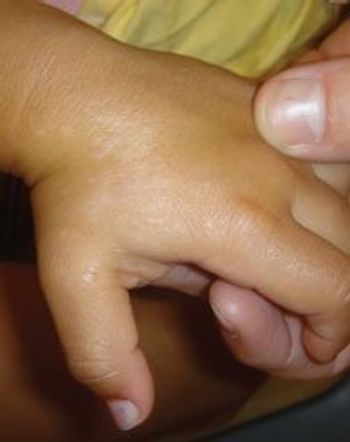
Linear bands of hypopigmentation on a child's hand.
Chances are you've seen its handiwork. It's molluscum contagiosum, and while many 'peds' choose to let this infection run its course, there can be benefits to intervention.
You are asked to evaluate a 2 ;½-year-old healthy boy with a 2-day history of a generalized asymptomatic skin eruption.
This baby boy was born at term to an 18-year-old primigravida via spontaneous vaginal delivery. The membranes ruptured about 6 hours before delivery. The amniotic fluid was heavily stained with meconium. Forceps were not used during the delivery. The newborn initially had poor tone and no spontaneous respirations, but his heart rate exceeded 100 beats per minute. Bulb and deep suctioning as well as supplemental oxygen were provided. Apgar scores were 3 and 8 at 1 and 5 minutes.
The parents of this 1-year-old girl brought her for evaluation of a neck mass of sudden onset (A). They had first noticed the mass on the morning of presentation. The child had cold symptoms and had been snoring, but she had no history of fever, shortness of breath, wheezing, or stridor. Her activity level and appetite had not changed.
A 4-year-old Hispanic boy was referred to our facility because of elevated levels of alanine trans- aminase (ALT) and aspartate transaminase (AST), which were detected during an evaluation of transient abdominal pain while the boy was in Puerto Rico. He was otherwise in perfect health; a review of systems was negative. His past medical history and birth history were noncontributory. Immunizations, including hepatitis B, were up-to-date. The family history was significant for tuberculosis and rheumatoid arthritis.
A 15-year-old girl was brought to the emergency department because of bilateral shoulder and hip pain associated with myalgia and fatigue. The symptoms had been present for 2 months and had increased in intensity over the past few days. The patient had systemic lupus erythematosus, asthma, and seizure disorder.
This rash on a teenage boy's palms began on his hands and spread to his torso and upper and lower extremities over several days. He had no pain or pruritus. Two weeks before the lesions appeared, he had experienced fatigue, fever, and myalgia of 1 week's duration.
This 6-year-old child was brought to the emergency department by her parents after these blisters suddenly appeared on her skin. She otherwise appeared to be well and was not febrile.
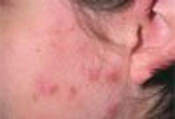
A 16-year-old girl is bothered by a spreading, itchy rash of 1 week’s duration on the left preauricular cheek. The otherwise healthy teenager denies taking any medications or using new cosmetics.
ABSTRACT: Because almost one tenth of American children aged 2 to 11 years have untreated tooth decay, a physical examination that includes inspection of the mouth is crucial. Look for cavitated or noncavitated lesions, dental fillings, and missing teeth; gingivitis and/or plaque, chalky white spots, or deep fissures on the teeth suggest dental decay. Dental care strategies that can be discussed at well-child visits include the benefits of daily flossing and brushing with fluoridated toothpaste, limited intake of dietary sugar, the establishment of a dental home, and use of protective mouthguards and face protectors during sport activities. Fluoride supplementation can be prescribed for children exposed to inadequate amounts in the water supply. The Caries-Risk Assessment Tool can help identify children at high risk for tooth decay. The pediatrician can have a great impact on ensuring that children obtain necessary dental care; a literature review found that children referred to a dentist by a primary care provider were more likely to visit a dentist than those not referred.
This lacy, purplish skin discoloration was noted on the trunk and lower extremities of a 6-week-old boy during a well-baby visit. The mother had noticed the discoloration periodically since birth. It was most apparent when the baby was cold.
Certain exanthems show a predilection for the summer. The path to diagnosing these conditions, however, can be anything but clear. A new algorithm sheds some much needed light.
A healthy 2-year-old was noted to have "blueberry" spots on her legs and trunk at birth.
A 9-month-old white boy was brought to the emergency department (ED) after he had been crying inconsolably for 2 hours. The parents thought the crying was related to the child's left leg, which they felt "did not look right." Two weeks earlier, the boy had a similar episode of inconsolable crying, and a fracture of the right distal radius was diagnosed. The fracture was presumptively caused by entrapment of his arm in the crib railings.
A 4-year-old boy presented with a pustular rash on the radial aspect of the right wrist. The rash appeared about 4 days earlier as a group of vesicles on an erythematous base. The lesions caused mild discomfort.
I agree with Dr Kirk Barber's comments about treating molluscum contagiosum: that removal of the lesion's core is curative and that autoinoculation at the time of treatment is only a small concern. I have found, however, that it is not necessary to go through the trouble of removing the core.
This infant was brought to the emergency department after a week-long exacerbation of her atopic eczema. She had not received any therapy for her eczema or for these erosions.
These 2 swellings on the scalp of a 13-year-old boy had appeared about 8 months earlier and had slowly enlarged. The mother had massaged the lesions when they were small in an attempt to heal them. There was no history of local trauma (eg, from hair-cutting tools) and no pain or discharge from the lesions.
An otherwise healthy 3-year-old girl was brought for evaluation of fever, sore throat, and shaking chills of 12 hours' duration. She had beefy-red, posterior oropharyngeal erythema and a scarlatiniform rash on her shoulder that had been present for a few hours. The rash faded out over her chest but reappeared in the perineum and lower abdomen. She also had vulvovaginal inflammation with surrounding erythema. The mother was surprised to see the inflammation, but in retrospect added that the child had complained of vaginal discomfort as well. A rapid antigen test for group A b-hemolytic streptococci (GABHS) from a swab of the oropharynx was positive.
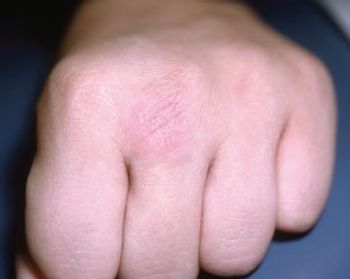
A 6-year-old girl is brought for evaluation of an asymptomatic indurated erythematous lesion that erupted on the metacarpophalangeal joint of one hand 2 weeks ago. The patient has no history of trauma or exposure and is otherwise healthy.
ABSTRACT: Dramatic progress has been made in our understanding of pediatric rheumatic disease. Various classification systems help identify juvenile idiopathic arthritis (JIA), which involves unique considerations that distinguish it from rheumatoid arthritis in adults. Vaccination issues are important for children with JIA. Renal involvement with systemic lupus erythematosus (SLE) is more common and more severe in children than in adults, but treatment of children who have SLE is similar to that of adults. Neonatal lupus erythematosus may occur in infants whose mothers have SLE. Juvenile dermatomyositis is associated with significant morbidity and mortality. Kawasaki disease is a common vasculitis of childhood, especially in infants and toddlers. Each of at least 8 major familial periodic fever clinical syndromes has specific distinguishing characteristics.
Advertisement
Advertisement
Trending on Contemporary Pediatrics
1
FDA approves acetaminophen-naproxen sodium combination tablet for ages 12 and older
2
US measles cases surpass 2025 total, hit highest level since 1991
3
Pooled phase 3 data show early, consistent tapinarof cream responses across pediatric age groups in AD
4
Ultra-processed foods tied to brain differences in young children
5