
An afebrile baby presents with disseminated pustules on the trunk, face, and extremities.
Dermatology
Latest News
Advertisement
Advertisement
A 17-month-old girl awoke with drooling, cough, respiratory distress, and a muffled cry and was brought to the emergency department. She had no nausea, vomiting, or diarrhea and no history of choking, aspiration, or airway problems.
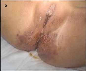
An 8-year-old girl is brought to the emergency department by her mother with a complaint of 5 days of worsening constipation and rectal bleeding. For the past week, the girl has had small stools 3 or 4 times a day and crampy abdominal pain. Yesterday, her stools were streaked with a small amount of blood. The mother notes that her daughter spends up to an hour in the bathroom with each bowel movement. In addition, the mother remarks that the girl has a rash in the rectal area and along the inner thighs.
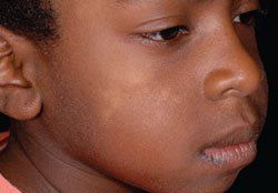
An 11-year-old girl presented with a swelling on the left side of the chin of 1 month's duration; in the past 24 hours, following a bite by an unidentified insect, the swelling had rapidly enlarged and become painful (A). She was otherwise healthy and had no significant medical or family history.
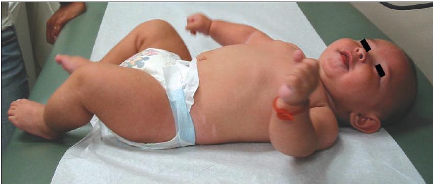
Numerous brown macules were sparsely distributed over the torso, head, and extremities of an African American newborn. The infant also had a mongolian spot on the buttocks. The baby's mother, a great aunt on the father's side, and the great aunt's daughter and grandson had similar brown macules at birth. All family members were healthy.
A 4-month-old infant is brought in due to raised hyperpigmented lesions around his shins.

For 2 days, a 17-year-old boy had a widespread pruritic eruption that involved the trunk and extremities but spared most of the face. Many of the lesions were annular, and they would appear and resolve within 1 day. The patient denied shortness of breath, difficulty in swallowing, and periorbital swelling.
Practicing pediatricians commonlytalk with patients and parentsabout medical risks. Examples of suchrisks include those of a newborn havinga genetic disease, of a complication of anillness developing, and of a patient experiencingan adverse effect from a medicationor vaccine. Different ways of expressingand communicating risk mayhelp patients and parents understand themagnitude of a risk and make informed,thoughtful decisions about their medicalcare. It is important to be aware of theinfluence personal experience and concernshave on how risk is perceived andto recognize how the choice of a particularway of framing a risk may inadvertentlycommunicate a clinician's personalbiases in a situation.
For the past few weeks, a 10-year-old boy had a pruritic abdominal rash that had not responded to over-the-counter topical medications. The rash had appeared around the time he started wearing a new belt (shown). The child was otherwise healthy. There was a family history of asthma.
In the newborn nursery, pediatricians commonly encounter infants born to mothers who were receiving selective serotonin reuptake inhibitors (SSRIs) for depression during pregnancy. Earlier studies suggested a number of potential effects of maternal SSRI use on the newborn; these included jitteriness, agitation, diarrhea, hypoglycemia, vomiting, hypothermia, respiratory distress, seizures, feeding difficulties, increased or decreased tone, low birth weight, and small size for gestational age.1
It is estimated that 12% of clinical methicillin-resistant Staphylococcus aureus (MRSA) infections are now community- associated.1 To combat the rising incidence of such infections, last month the CDC launched a new national campaign.
A previously healthy 16-month-old boy was hospitalized because of vomiting of 10 days' duration, fever of 4 days' duration (temperature up to 38.6°C [101.4°F]), and watery diarrhea. He also had had a maculopapular rash, which resolved the day before presentation. Family history was unremarkable.
The cause was pilonidal sinus disease. The term pilonidal is derived from the Latin words "pilus," meaning "hair," and "nidus," meaning "nest." Pilonidal sinus disease is more common in males than in females and typically appears during adolescence. About 1% of all males and 0.1% of all females have an asymptomatic pilonidal sinus with potential for disease.1 The suspected overall incidence is about 1 in 5000. The disease seems to be most prevalent in those with stiff, dark or auburn hair, although it has been observed in all races.2
Parents are understandably concerned about vaccines and autism, a relationship publicized by lawsuits, alternative therapies, and claims of government cover-up.
A healthy 10-year-old girl has had a red, white, and blue lump on her thigh since birth.
Many factors can be considered in attempting to establish the cause of a skin disorder. These include the color, morphology, and location of the lesions; associated symptoms, such as itching and fever; and exposure to drugs or to other children who have a rash. Linearity of the lesions may also suggest the diagnosis.
In addition to syringohydromelia and meningocele, the MRI of the spine showed a fluid-filled mllerian duct remnant that extended from the base of the bladder to the posterior superior aspect of the prostate gland. The margins of the fluid collection in the remnant are smoothly bound by a hypointense structure that represents a discrete tissue wall. A mllerian duct remnant can be confused with free fluid in the cul-de-sac posterior to the bladder.
A 4-year-old boy was brought to the hospital because of fever (temperature of 39.4°C [103°F]) and a bright, salmon-pink rash on the palms and soles that was associated with peeling (A and B). His symptoms had been present for 2 days. During that time, he also had redness of the eyes, fatigue, and anorexia.
There is increasing evidence that the inflammatory nature of psoriasis is associated with an increase in comorbid conditions, such as obesity and cardiovascular disease, and that people with psoriasis have a shortened life expectancy.
A discussion of two types of Genital Lesions: Smegma and Torsion of the Testis.
Hemangiomas are the most common tumors seen in infants. Despite their rapid growth early in life, often the only action required of the physician is to reassure the parents that such tumors usually involute spontaneously during childhood.
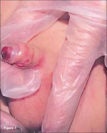
A 7-month-old male infant was brought to the emergency department (ED) by his biological mother, who reported noticing dried blood on the baby's penis and in his mouth. For several hours prior, he had been in the care of her boyfriend. On physical examination, there were severe ecchymoses and petechiae on the penile glans and shaft (Figure 1), ecchymoses on the right side of the soft palate, a laceration of the lingular frenulum, and a 2-cm bruise with dried blood over the right lip.
A 7-year-old boy presented to his primary care pediatrician with a 24-hour history of vomiting, abdominal pain, and low-grade fever. The child appeared stable. A viral illness was diagnosed. The child was sent home, and his parents were advised to give him adequate fluids as well as acetaminophen as needed for fever.
Sixteen-year-old African American girl with pruritic, painful lesions on both thighs that progressively worsened over 2 weeks and later spread to the trunk and upper and lower extremities.
A 3-month-old boy was brought to the pediatrician with a 2-day history of "moaning," lethargy, and difficulty in latching on for breast-feeding. The infant had not had a wet diaper for the past 24 hours, and his last bowel movement was more than 48 hours ago.
Advertisement
Advertisement
Trending on Contemporary Pediatrics
1
FDA approves acetaminophen-naproxen sodium combination tablet for ages 12 and older
2
US measles cases surpass 2025 total, hit highest level since 1991
3
Pooled phase 3 data show early, consistent tapinarof cream responses across pediatric age groups in AD
4
Prenatal therapy improves outcomes in fetal renal failure
5