A 12-year-old girl presents with a 1-year history of itchy lesions on her right leg. The lesions are worse during the winter. She has a history of atopic dermatitis and asthma.
Alexander K. C. Leung, MD
Advertisement
Articles by Alexander K. C. Leung, MD
Patient delivered vaginally at term to a G2P1 24-year-old mother following an uncomplicated pregnancy. Apgar scores, 7 and 9 at 1 and 5 minutes, respectively. No history of maternal exposure to teratogens. Parents non-consanguineous. No family history of congenital or chromosomal abnormality.
Ten-day-old boy born vaginally at 37 weeks breech without complications. Has history of poor feeding with vomiting and has lost weight since birth. One episode of vomiting described as projectile. Ultrasonography ruled out pyloric stenosis but revealed bilateral hydronephrosis. Patient referred to the emergency department for further evaluation.
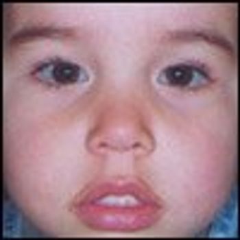
his 7-year-old boy presented with mental retardation and delayed gross motor milestones: he first sat up at 12 months, walked at 18 months, and ran at 2 years. His level of speech development was that of a 5-year-old; his IQ was 75.
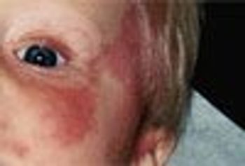
Sixteen-year-old with a recurrent, painful, pruritic rash on right cheek and right eyelid. Current outbreak started 2 days earlier. The rash always appears in the same fashion and in the same location; it typically lasts a few days and resolves spontaneously.
Photo Essay: A Collage of Infectious Diseases in Children Perichondritis Periorbital Cellulitis Bacterial Conjunctivitis Roseola Infantum (Exanthem Subitum) Tinea Corporis ("Ringworm")
Seven-month-old boy with hyperpigmented lesions on his legs. Lesions first noted at 4 months of age. His 32-year-old father and 58-year-old grandfather had similar lesions on their legs.

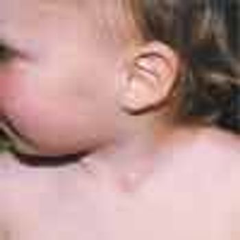
Six-year-old East Indian boy delivered by normal spontaneous vaginal delivery to a para 3, gravida 2, 42-year-old mother following uncomplicated, full-term pregnancy. Apgar scores: 9 and 9, at 1 and 5 minutes, respectively. Birth weight, 2.5 kg. Infant hypotonic at birth with numerous dysmorphic features. Delayed developmental milestones; IQ measured at 80.
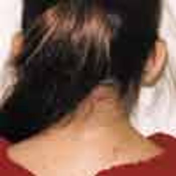
Ten-year-old girl assessed because of limitation of neck motion. Born at term to non-consanguineous parents. No history of necktrauma. Family history andpast health unremarkable.
The April issue of Consultant For Pediatricians included a case of a 12-year-old girl with poliosis. The author, Bhagwan Das Bang, MD, noted that poliosis is associated with ocular chronic staphylococcal blepharitis, Waardenburg syndrome, Marfan syndrome, vitiligo, and Vogt-Koyanagi syndrome.

Seven-year-old boy with red, nonpruritic rash that appeared first on the cheeks and then spread to the trunk, extremities, and buttocks. No history of respiratory, GI, or other symptoms in the several weeks before the onset of the rash. Patient is otherwise healthy.
A 7-year-old boy presented with an asymptomatic cystic lesion on the lateral aspect of the left ankle of 4 months' duration. There was no history of trauma. The mass fluctuated in size: it was smaller when the child was recumbent and larger when the child was upright.
A 9-year-old girl presents with multiple hyperpigmented lesions, some of which have been present since birth (Figure 1). The lesions have increased in size and number. There is no history of seizures. Her 40-year-old mother has multiple skin nodules (Figure 2).
A mother brought in her 3-week-old son on the day she discovered a reddish urine stain in the baby's diaper. There was no stool in the diaper. The boy had been circumcised on the second day of life, and the mother was concerned that her son might have experienced a complication of the procedure.
A 12-year-old boy is assessed on the same day that he passed red-colored urine. The boy had been vigorously wrestling with his older brother in the morning; he passed the abnormal-colored urine after lunch. His mother was worried that his kidneys might have been injured during the wrestling.
An 8-year-old boy is assessed on the same day that he passed red-colored urine. The boy first noted the abnormal-colored urine when he voided on awakening.
Key words: type 2 diabetes melllitus, childhood obesity
An 18-year-old girl presented with an asymptomatic nodule on the posterior aspect of the right upper arm. The lesion had developed a month after an episode of chickenpox at 11 years of age and had slowly enlarged. The lesion was 7 mm in diameter; it was firm, rubbery, reddish brown, and nontender.
A 10-month-old girl with a mass on left side of neck. Swelling first noticed by mother when child was 7 months old. The infant is eating well, thriving, and otherwise healthy.
This black lesion had been present on the upper back of a 5-year-old girl since her birth. The lesion had gradually enlarged to its current size of 1.5 cm. In the past year, 3 satellite black macules had developed in the surrounding area.

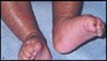
Female infant born to a gravida II, para I, 23-year-old mother at 38 weeks' gestation. Pregnancy complicated by oligohydramnios. Cesarean delivery performed because of prolonged time after rupture of membranes and fetal distress. Apgar scores, 3 and 6 at 1 minute and 5 minutes, respectively.
This 14-year-old girl had first noticed the well-circumscribed, roughened, irregular growth on her right middle finger 6 months earlier. The lesion had progressively enlarged. The girl's mother had a similar, but smaller, lesion on the left elbow. Alexander K. C. Leung, MD, and Justine H. S. Fong, MD, of Calgary, Alberta, diagnosed verruca vulgaris. This proliferative, hyperkeratotic, exophytic lesion is most commonly caused by human papillomavirus types 2 and 4.
A 3-year-old boy who presents with blue sclerae and a history of tibial fracture following a minor trau- ma (jump from a height of less than 18 inches). Has a long-standing complaint of back pain. Mother remarks that the boy bruises easily. Medical history otherwise unremarkable.
A 13-year-old boy presented with an explosive eruption of numerous, small, round, erythematous, itchy plaques on his lower back and lower limbs of 2 weeks' duration (A). Some of the lesions were scaly. His nails were normal. There was no evidence of arthritis or joint deformity. He had a sore throat a month before the onset of the rash but did not seek medical attention. He was not taking any medication and had no history of joint pain or family history of skin problems.
The mother of this 3-month-old girl was concerned about her baby's diffuse, gradual loss of scalp hair. The infant was otherwise healthy and was feeding normally.
Photo Essay: Factitious Dermatitis Lip Licker's Dermatitis Ecchymoses From Spoon Scratching Ecchymosis From Cupping
For the past 10 days, a 3-week-old infant had a rash on the face. He was born at term to a healthy, 22-year-old primigravida, following an uncomplicated pregnancy and normal spontaneous vaginal delivery (birth weight, 3.1 kg; length, 49.5 cm). Numerous comedones and papules were noted on the infant's cheeks.
A 10-month-old infant was referred for evaluation of possible Sturge-Weber syndrome. According to his parents, the discoloration on the child’s face was present at birth. Physical examination revealed an otherwise healthy infant with extensive port-wine stains on his face. Ophthalmologic and neurologic examination findings were normal.
Advertisement
Advertisement
Advertisement
Trending on Contemporary Pediatrics
1
Cyclosporiasis in 2026: A clinical update for pediatricians
2
Nonreceipt of newborn intramuscular vitamin K linked to doubled bleeding risk
3
Udenafil study shows lower ELF scores in Fontan-associated liver disease
4
Supporting the language development of deaf and hard of hearing children: A clinical guide for pediatric providers
5