


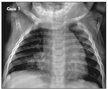
A 6-month-old boy with 1-week history of dry cough that worsened at night. He had been wheezing off and on for the past month and had visited the emergency department on one occasion.
A 6-month-old boy with 1-week history of dry cough that worsened at night. He had been wheezing off and on for the past month and had visited the emergency department on one occasion.

In this podcast, Dr John Kelso dispels myths and makes a case for office-based spirometry for pediatricians.
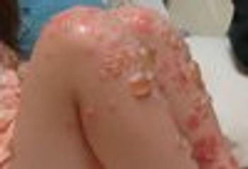
A 4-year-old, previously healthy girl presented to a tertiary care children’s hospital emergency department (ED) with large, tense bullae involving up to 40% of her body surface area. The patient’s parents described a 12-day history of itchy, papulovesicular lesions that had progressed into large blisters.

As the calendar advances to the fall months, we are reminded everywhere that it’s time for the annual influenza vaccination. In August 2011, the Advisory Committee on Immunization Practices (ACIP) updated its vaccination recommendations to include new guidelines for children ages 6 months through 8 years, and for persons with egg allergy.

Like many physicians, I find it difficult to care for a patient with a terminal disease-a discomfort heightened when the patient is an ebullient, beautiful child.

My 18-month-old son has had a diaper rash, with no other symptoms, within a few days of eating a poached egg on 3 separate occasions.
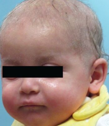
This 9–month–old boy was initially evaluated at age 6 weeks for an extensive eczematous rash on the head and antecubital and diaper areas and blood and mucus in the stool with each diaper change over a 2– to 3–week period. The symptoms were attributed to milk allergy, and the infant’s formula was changed. At 8 weeks of age, a petechial rash developed on the boy’s trunk and legs. His symptoms persisted despite multiple formula changes, and he was referred to the emergency department.
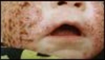
A 7-month-old boy with a history of severe atopic dermatitis and asthma was brought for evaluation of a generalized rash, fever, and irritability of 2 days’ duration. He had no respiratory symptoms. His medical history was significant for anorexia, without vomiting or diarrhea. He had a strong family history of allergy.
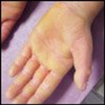
At his first well-child visit after a family move, an 8-year-old boy was noted to have bilateral erythematous plaques on the surfaces of his hands and feet. Mother reported that the condition had been present since he was 2 or 3 months old. Patient’s father and other male relatives on the paternal side (uncles, grandfather, great-grandfather) were similarly affected. No other associated symptoms, such as hyperhidrosis, reported. The child did not have a history of eczema, asthma, or food allergies; however, he did have a history of allergic rhinitis and occasional pruritus.
“Drug rash” is a common pediatric complaint in both inpatient and outpatient settings. This term, however, denotes a clinical category and is not a precise diagnosis. Proper identification and classification of drug eruptions in children are important for determining the possibility of-and preventing progression to-internal involvement. Accurate identification is also important so that patients and their parents can be counseled to avoid future problematic drug exposures.
Match the photographs of the rashes with the correct diagnosis.
Allergy testing can aid the diagnosis of allergic disorders; however, it is not diagnostic. With skin testing, in particular, a positive result does not necessarily indicate clinical allergy, and a negative result does not always exclude clinical relevance.
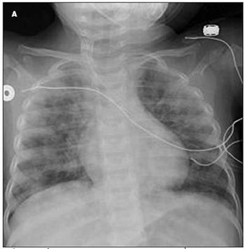
A 16-month-old girl was initially brought to her primary care physician because of persistent nonproductive cough of 1 to 2 weeks’ duration, with lethargy, poor feeding, and worsening cough for the past 36 hours. She had been afebrile. The patient was noted to be pale and had a decreased level of interaction. She was promptly sent to the local emergency department. Laboratory studies showed a hemoglobin level of 3.8 g/dL and hypochromic microcytic anemia. The patient was subsequently transferred to the pediatric ICU for evaluation.
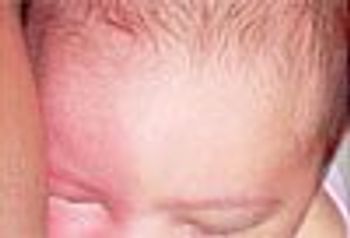
Episodic right-sided facial flushing was noted in a 2-month-old girl born at full term via forceps-assisted vaginal delivery. The erythema appeared within minutes of latching onto her mother’s breast and resolved within 5 to 10 minutes after breastfeeding. The episodes of flushing had begun a week before the clinic visit; there were no collateral symptoms of anaphylaxis. Because food allergy was suspected, the mother had eliminated all dairy products from her diet.
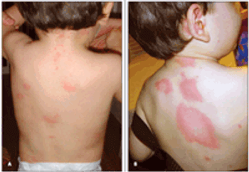
Circumscribed erythematous lesions developed on the back and abdomen of this 19-month-old boy. The rash was mildly pruritic. The parents gave the child 1 dose of diphenhydramine, and the rash resolved after an hour. About 12 hours later, new lesions developed on the face, neck, and upper back. The child was given the same treatment and the symptoms resolved. The following morning, widespread lesions were noted on the child's face, neck, trunk, and extremities.
A 5-month-old Asian boy was brought for evaluation of hair loss and a red, scaly rash on the scalp and body. The rash had not responded to hydrocortisone 2.5% ointment. There was a family history of asthma, food allergies, and allergic rhinitis. His mother had Hashimoto thyroiditis.
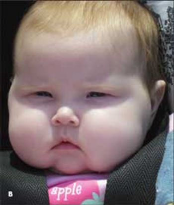
A 5-month-old girl with progressively worsening generalized rash of 3 weeks’ duration. No obvious sensitivity, fever, recent infection, medication use, or known contact with irritant.
For 2 days, a 17-year-old boy had a widespread pruritic eruption that involved the trunk and extremities but spared most of the face. Many of the lesions were annular, and they would appear and resolve within 1 day. The patient denied shortness of breath, difficulty in swallowing, and periorbital swelling.
A 7-year-old boy with annular, asymptomatic, flesh-colored lesion onthe wrist that had developed slowly over the past month. The parents hadremoved the child from school because they were told that the lesion wasringworm. The lesion had failed to resolve after application of an antifungalcream for 10 days.
A 4-year-old boy presented for further evaluation of persistent right hip painof 2 months’ duration. Before the onset of the pain, he had been limping,favoring his right side. For several days before he was brought in forevaluation, he had had fevers and sweating in addition to the right hippain.
I enjoyed Dr Kirk Barber’s interesting Dermclinic quiz featuring a 5-year-old boy with a dramatic resentation of tinea corporis (CONSULTANT FOR PEDIATRICIANS, February 2009, page 43). I am curious to know why Dr Barber prescribed oral terbinafine for this patient.
A persistent, eczematous dermatitis had developed in the perioral area during the winter months in this 10-year-old boy. Topical corticosteroid creams had been tried, and these seemed to help some, but the ondition never really cleared. Because of the failure of the corticosteroid creams, a topical antifungal cream had also been tried; however, this, too, was of limited effectiveness.
An 18-month-old white boy is brought to his well-care visit by his parents, who are concerned that for the past month he has been less social and active. He has appeared weak and has refused to walk or play with his siblings. He has also had a decreased appetite and has lost about 2 kg. He has vomited several times but with no bile or blood.
The most common chronic medical problem that we pediatricians treat is asthma. We do our best to manage our patients’ asthma by prescribing controller medications, providing asthma action plans, and guiding families through acute exacerbations. We often ask about possible environmental triggers, such as tobacco smoke and cockroaches, and we advise patients to reduce their exposure to those triggers.
The most common chronic medical problem that we pediatricians treat is asthma. We do our best to manage our patients' asthma by prescribing controller medications, providing asthma action plans, and guiding families through acute exacerbations. We often ask about possible environmental triggers, such as tobacco smoke and cockroaches, and we advise patients to reduce their exposure to those triggers.